Application
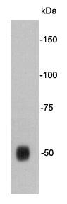
Fig1: Western blot analysis of Tau on human brain tissue lysate using anti-Tau antibody at 1/1,000 dilution.Application
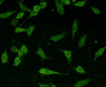
Fig2: ICC staining Tau in SHG-44 cells (green). The nuclear counter stain is DAPI (blue). Cells were fixed in paraformaldehyde, permeabilised with 0.25% Triton X100/PBS.Application
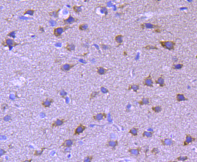
Fig3: Immunohistochemical analysis of paraffin-embedded rat brain tissue using anti-Tau antibody. Counter stained with hematoxylin.Application
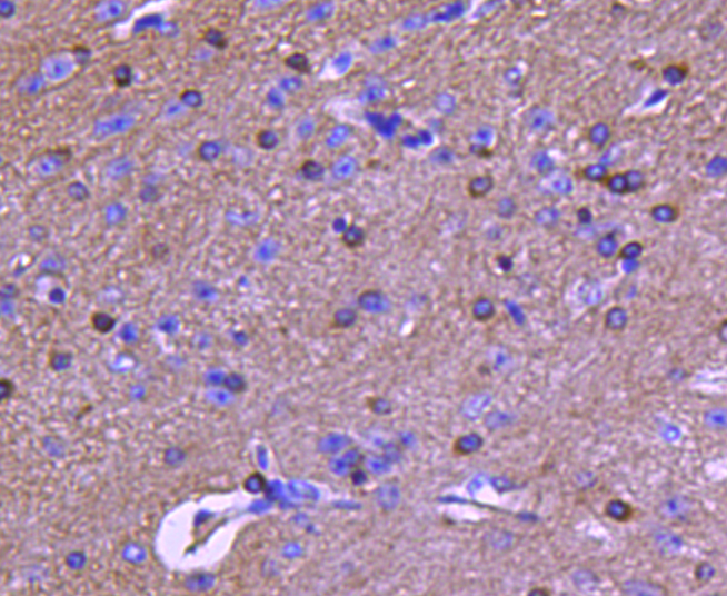
Fig4: Immunohistochemical analysis of paraffin-embedded mouse brain tissue using anti-Tau antibody. Counter stained with hematoxylin.| Product Name | Tau |
|---|---|
| Antibody Type | Primary Antibodies |
| Antigen Alias | AI413597 antibody AW045860 antibody DDPAC antibody FLJ31424 antibody FTDP 17 antibody G protein beta1/gamma2 subunit interacting factor 1 antibody MAPT antibody MAPTL antibody MGC134287 antibody MGC138549 antibody MGC156663 antibody Microtubule associated protein tau antibody Microtubule associated protein tau isoform 4 antibody Microtubule-associated protein tau antibody MSTD antibody Mtapt antibody MTBT1 antibody MTBT2 antibody Neurofibrillary tangle protein antibody Paired helical filament tau antibody Paired helical filament-tau antibody PHF tau antibody PHF-tau antibody PPND antibody PPP1R103 antibody Protein phosphatase 1, regulatory subunit 103 antibody pTau antibody RNPTAU antibody TAU antibody TAU_HUMAN antibody Tauopathy and respiratory failure, included antibody |
| Product description | Tau protein is a highly soluble microtubule-associated protein (MAP). In humans, these proteins are found mostly in neurons compared to non-neuronal cells. One of tau's main functions is to modulate the stability of axonal microtubules. Other nervous system MAPs may perform similar functions, as suggested by tau knockout mice that did not show abnormalities in brain development - possibly because of compensation in tau deficiency by other MAPs. Six tau isoforms exist in human brain tissue, and they are distinguished by their number of binding domains. Three isoforms have three binding domains and the other three have four binding domains. Tau is a phosphoprotein with 79 potential Serine (Ser) and Threonine (Thr) phosphorylation sites on the longest tau isoform. Phosphorylation has been reported on approximately 30 of these sites in normal tau proteins. Hyperphosphorylation of the tau protein (tau inclusions, pTau) can result in the self-assembly of tangles of paired helical filaments and straight filaments, which are involved in the pathogenesis of Alzheimer's disease and other tauopathies. |
| Immunogen | Synthetic peptide (KLH-coupled) within human Tau N-terminal. |
| Clonality | Polyclonal |
|---|---|
| Isotype | IgG |
| Host Species | Rabbit |
| Tested Applications | WBICCIHC |
| WB:1:500-1:2,000 ICC:1:50-1:200 IHC:1:50-1:200 |
|
| Species Reactivity | HumanMouseRat |
| Concentration | 1mg/ml |
| Alternative Names | AI413597 antibody
AW045860 antibody
DDPAC antibody
FLJ31424 antibody
FTDP 17 antibody
G protein beta1/gamma2 subunit interacting factor 1 antibody
MAPT antibody
MAPTL antibody
MGC134287 antibody
MGC138549 antibody
MGC156663 antibody
Microtubule associated protein tau antibody
Microtubule associated protein tau isoform 4 antibody
Microtubule-associated protein tau antibody
MSTD antibody
Mtapt antibody
MTBT1 antibody
MTBT2 antibody
Neurofibrillary tangle protein antibody
Paired helical filament tau antibody
Paired helical filament-tau antibody
PHF tau antibody
PHF-tau antibody
PPND antibody
PPP1R103 antibody
Protein phosphatase 1 regulatory subunit 103 antibody pTau antibody RNPTAU antibody TAU antibody TAU_HUMAN antibody Tauopathy and respiratory failure included antibody |
|---|---|
| Molecular Weight(MW) | 50kDa |
| Cellular Localization | Cytoplasm |
Application
Fig1: Western blot analysis of Tau on human brain tissue lysate using anti-Tau antibody at 1/1,000 dilution.
Application
Fig2: ICC staining Tau in SHG-44 cells (green). The nuclear counter stain is DAPI (blue). Cells were fixed in paraformaldehyde, permeabilised with 0.25% Triton X100/PBS.
Application
Fig3: Immunohistochemical analysis of paraffin-embedded rat brain tissue using anti-Tau antibody. Counter stained with hematoxylin.
Application
Fig4: Immunohistochemical analysis of paraffin-embedded mouse brain tissue using anti-Tau antibody. Counter stained with hematoxylin.| Positive Control | Human brain tissue, SHG-44, rat brain tissue, mouse brain tissue. |
|---|---|
| Application Notes | WB:1:500-1:2,000 ICC:1:50-1:200 IHC:1:50-1:200 |
| Form | Liquid |
|---|---|
| Storage Instructions | Store at +4℃ after thawing. Aliquot store at -20℃. Avoid repeated freeze / thaw cycles. |
| Storage Buffer | 1*TBS (pH7.4), 1%BSA, 40%Glycerol. Preservative: 0.05% Sodium Azide. |


