Application
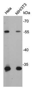
Fig1: Western blot analysis on cell lysates using anti-GLUT4 rabbit polyclonal antibodies.Application
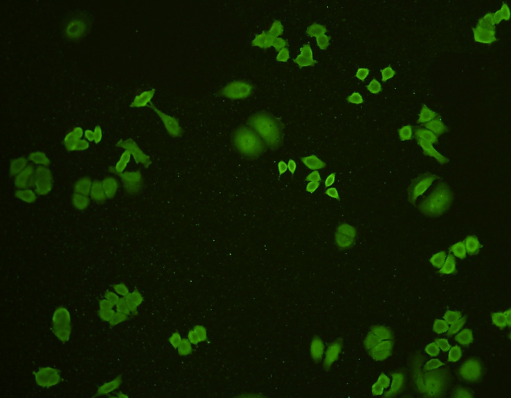
Fig2: ICC staining GLUT4 in MCF-7 cells (green). Cells were fixed in paraformaldehyde, permeabilised with 0.25% Triton X100/PBS.Application
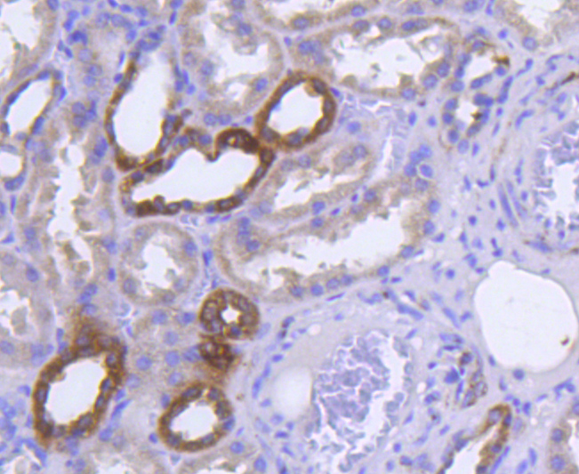
Fig3: Immunohistochemical analysis of paraffin-embedded human kidney tissue using anti-GLUT4 antibody. Counter stained with hematoxylin.Application
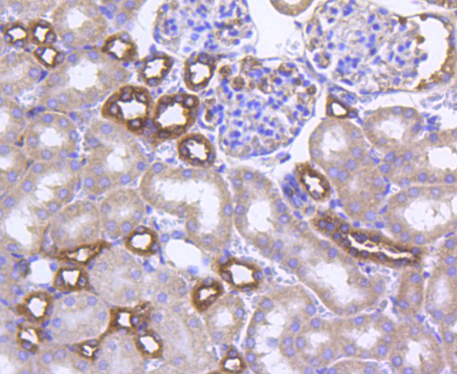
Fig4: Immunohistochemical analysis of paraffin-embedded mouse kidney tissue using anti-GLUT4 antibody. Counter stained with hematoxylin.Application
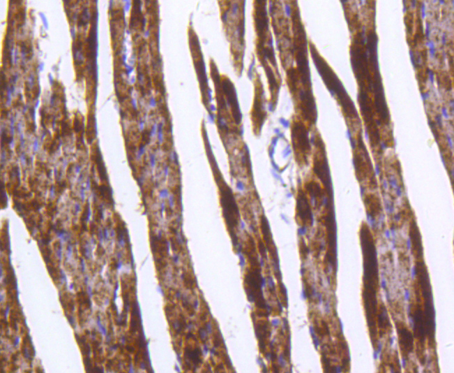
Fig5: Immunohistochemical analysis of paraffin-embedded mouse heart tissue using anti-GLUT4 antibody. Counter stained with hematoxylin.| Product Name | Glucose Transporter GLUT4 |
|---|---|
| Antibody Type | Primary Antibodies |
| Antigen Alias | Glucose transporter GLUT 4 Glucose transporter type 4 Glucose transporter type 4 insulin responsive GLUT 4 GLUT-4 GLUT4 GTR4_HUMAN Insulin responsive glucose transporter type 4 insulin-responsive kug SLC 2A4 SLC2A4 solute carrier family 2 (facilitated glucose transporter) member 4 Solute carrier family 2 member 4 Solute carrier family 2, facilitated glucose transporter member 4 |
| Product description | GLUT4 is the insulin-regulated glucose transporter found primarily in adipose tissues and striated muscle (skeletal and cardiac) that is responsible for insulin-regulated glucose transport into the cell. Under conditions of low insulin, GLUT4 is sequestered in intracellular vesicles in muscle and fat cells. Insulin induces a rapid increase in the uptake of glucose by inducing the translocation of GLUT4 from these vesicles to the plasma membrane. Muscle contraction stimulates muscle cells to translocate GLUT4 receptors to their surfaces. This is especially true in cardiac muscle, where continuous contraction can be relied upon; but is observed to a lesser extent in skeletal muscle. |
| Immunogen | peptide |
| Clonality | Polyclonal |
|---|---|
| Isotype | IgG |
| Host Species | Rabbit |
| Tested Applications | WBICC/IFIHC |
| WB:1:500~1:2000 ICC/IF:1:200~1:500 IHC:1:50~1:200 Notes:Optimal dilutions/concentrations should be determined by the researcher. |
|
| Species Reactivity | HumanMouse |
| Concentration | 1mg/ml |
| Alternative Names | Glucose transporter GLUT 4
Glucose transporter type 4
Glucose transporter type 4 insulin responsive
GLUT 4
GLUT-4
GLUT4
GTR4_HUMAN
Insulin responsive glucose transporter type 4
insulin-responsive
kug
SLC 2A4
SLC2A4
solute carrier family 2 (facilitated glucose transporter) member 4
Solute carrier family 2 member 4
Solute carrier family 2 facilitated glucose transporter member 4 |
|---|---|
| Molecular Weight(MW) | 55 kDa |
| Cellular Localization | Cell membrane |
| SwissProt ID | P14672 |
|---|
Application
Fig1: Western blot analysis on cell lysates using anti-GLUT4 rabbit polyclonal antibodies.
Application
Fig2: ICC staining GLUT4 in MCF-7 cells (green). Cells were fixed in paraformaldehyde, permeabilised with 0.25% Triton X100/PBS.
Application
Fig3: Immunohistochemical analysis of paraffin-embedded human kidney tissue using anti-GLUT4 antibody. Counter stained with hematoxylin.
Application
Fig4: Immunohistochemical analysis of paraffin-embedded mouse kidney tissue using anti-GLUT4 antibody. Counter stained with hematoxylin.
Application
Fig5: Immunohistochemical analysis of paraffin-embedded mouse heart tissue using anti-GLUT4 antibody. Counter stained with hematoxylin.| Positive Control | Hela, MCF-7, NIH/3T3 |
|---|---|
| Application Notes | WB:1:500~1:2000 ICC/IF:1:200~1:500 IHC:1:50~1:200 Notes:Optimal dilutions/concentrations should be determined by the researcher. |
| Form | Liquid |
|---|---|
| Storage Instructions | Store at +4℃ after thawing. Aliquot store at -20℃ or -80℃. Avoid repeated freeze / thaw cycles. |
| Storage Buffer | 1*TBS (pH7.4), 0.5%BSA, 40%Glycerol. Preservative: 0.05% Sodium Azide. |


