Application
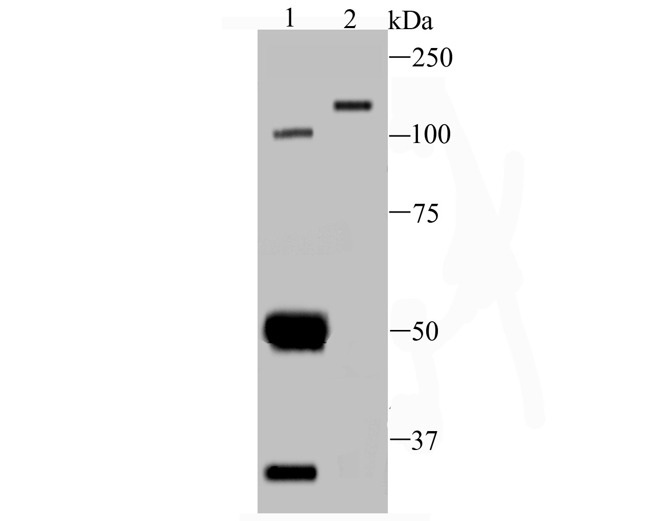
Fig1: Western blot analysis of AXL on different lysates using anti-AXL antibody at 1/100 dilution.Positive control:Lane1: Mouse kidney tissueLane2: CRCApplication
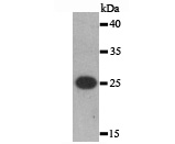
Fig2: Western blot analysis of AXL on human AXL recombinant protein using anti-AXL antibody at 1/1,000 dilution.Application
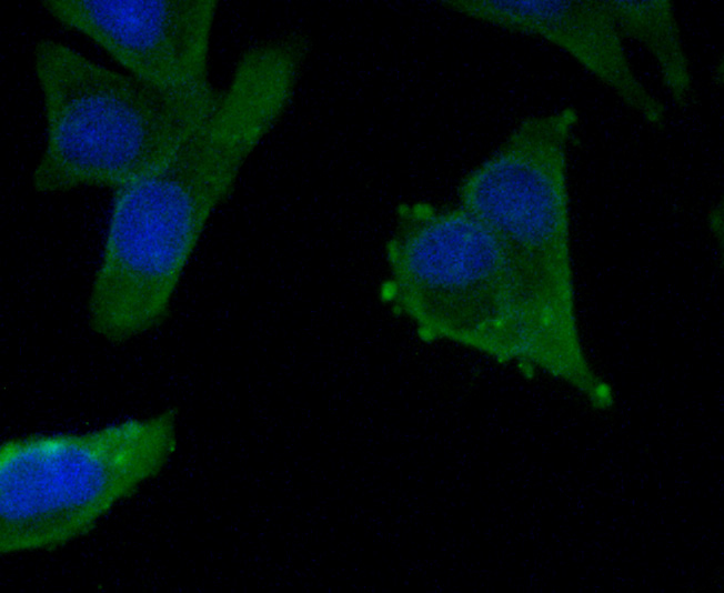
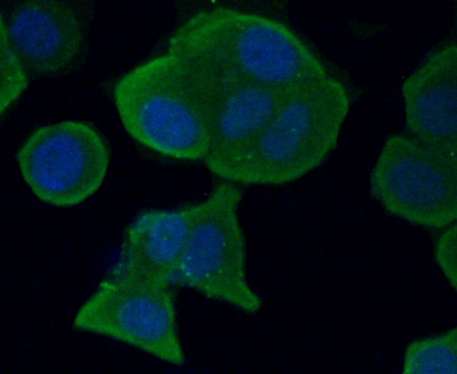
Fig3: ICC staining AXL in Hela cells (green). The nuclear counter stain is DAPI (blue). Cells were fixed in paraformaldehyde, permeabilised with 0.25% Triton X100/PBS.Application
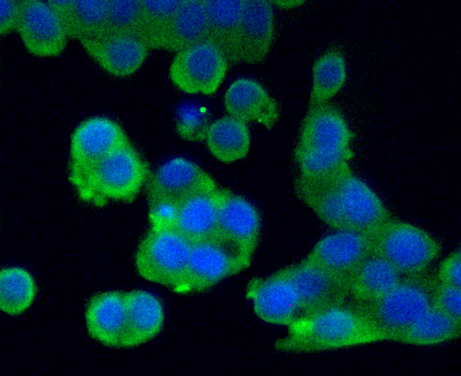
Fig4: ICC staining AXL in LOVO cells (green). The nuclear counter stain is DAPI (blue). Cells were fixed in paraformaldehyde, permeabilised with 0.25% Triton X100/PBS.Application
Fig5: ICC staining AXL in MCF-7 cells (green). The nuclear counter stain is DAPI (blue). Cells were fixed in paraformaldehyde, permeabilised with 0.25% Triton X100/PBS.| Product Name | AXL |
|---|---|
| Antibody Type | Primary Antibodies |
| Antigen Alias | Adhesion related kinase antibody AI323647 antibody Ark antibody Axl antibody AXL oncogene antibody AXL receptor tyrosine kinase antibody AXL transforming gene antibody AXL transforming sequence/gene antibody EC 2.7.10.1 antibody JTK11 antibody Oncogene AXL antibody Tyro7 antibody Tyrosine protein kinase receptor UFO antibody Tyrosine-protein kinase receptor UFO antibody UFO antibody UFO_HUMAN antibody |
| Product description | The UFO family of receptor tyrosine kinases is comprised of subfamily members Rse (also designated Tyro3, Sky, Brt, Dtk, Etk2 and Tif), Axl (also designated UFO or ARK) and Mer (also designated Nyk or Eyk). Rse is expressed Preferentially in the adult brain with lower expression in other tissues. Axl is found at highest levels in heart and skeletal muscle. Mer has been identified as a tyrosine kinase is involved in the development of glioblastomas. It is expressed at highest levels in ovary, prostate , Lung and kidney. Gas6, a growth arrest specific gene, and the related anticoagulation factor Protein S, have been identified as ligands for the UFO family of receptors. |
| Immunogen | Recombinant protein within human AXL 1-200 aa. |
| Clonality | Polyclonal |
|---|---|
| Isotype | IgG |
| Host Species | Rabbit |
| Tested Applications | WBICC |
| WB:1:500 ICC:1:50-1:200 |
|
| Species Reactivity | HumanMouse |
| Concentration | 2mg/ml |
| Alternative Names | Adhesion related kinase antibody AI323647 antibody Ark antibody Axl antibody AXL oncogene antibody AXL receptor tyrosine kinase antibody AXL transforming gene antibody AXL transforming sequence/gene antibody EC 2.7.10.1 antibody JTK11 antibody Oncogene AXL antibody Tyro7 antibody Tyrosine protein kinase receptor UFO antibody Tyrosine-protein kinase receptor UFO antibody UFO antibody UFO_HUMAN antibody |
|---|---|
| Molecular Weight(MW) | 98 kDa(Predicted band) |
| Cellular Localization | Membrane. |
Application
Fig1: Western blot analysis of AXL on different lysates using anti-AXL antibody at 1/100 dilution.Positive control:Lane1: Mouse kidney tissueLane2: CRC
Application
Fig2: Western blot analysis of AXL on human AXL recombinant protein using anti-AXL antibody at 1/1,000 dilution.
Application
Fig3: ICC staining AXL in Hela cells (green). The nuclear counter stain is DAPI (blue). Cells were fixed in paraformaldehyde, permeabilised with 0.25% Triton X100/PBS.
Application
Fig4: ICC staining AXL in LOVO cells (green). The nuclear counter stain is DAPI (blue). Cells were fixed in paraformaldehyde, permeabilised with 0.25% Triton X100/PBS.
Application
Fig5: ICC staining AXL in MCF-7 cells (green). The nuclear counter stain is DAPI (blue). Cells were fixed in paraformaldehyde, permeabilised with 0.25% Triton X100/PBS.| Positive Control | Mouse kidney tissue lysate, CRC cell lysate, human AXL recombinant protein lysate, Hela, LOVO, MCF-7. |
|---|---|
| Application Notes | WB:1:500 ICC:1:50-1:200 |
| Form | Liquid |
|---|---|
| Storage Instructions | Store at +4℃ after thawing. Aliquot store at -20℃ or -80℃. Avoid repeated freeze / thaw cycles. |
| Storage Buffer | 1*TBS (pH7.4), 1%BSA, 40%Glycerol. Preservative: 0.05% Sodium Azide. |


