Application
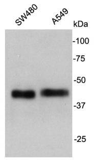
Fig1: Western blot analysis on cell lysates using anti- Cathepsin B rabbit polyclonal antibodies.Application
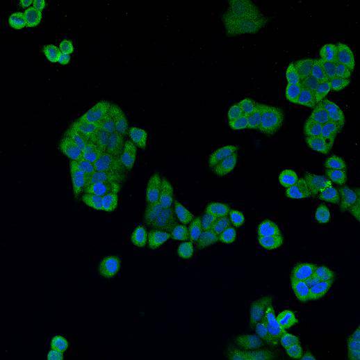
Fig2: ICC staining Cathepsin B in Hela cells (green). Cells were fixed in paraformaldehyde, permeabilised with 0.25% Triton X100/PBS and counterstained with DAPI in order to highlight the nucleus (blue).| Product Name | Cathepsin B |
|---|---|
| Antibody Type | Primary Antibodies |
| Antigen Alias | Amyloid precursor protein secretase antibody APP secretase antibody APPS antibody CATB_HUMAN antibody Cathepsin B heavy chain antibody Cathepsin B1 antibody CathepsinB antibody CPSB antibody CTSB antibody cysteine protease antibody OTTHUMP00000116009 antibody OTTHUMP00000229510 antibody OTTHUMP00000229511 antibody OTTHUMP00000229512 antibody OTTHUMP00000229514 antibody OTTHUMP00000229515 antibody OTTHUMP00000229516 antibody Preprocathepsin B antibody |
| Product description | Cathepsin B is an enzymatic protein belonging to the peptidase (or protease) families. In humans, it is coded by the CTSB gene. A wide array of diseases results in elevated levels of cathepsin B, which causes numerous pathological processes including cell death, inflammation, and production of toxic peptides. Focusing on neurological diseases, cathepsin B gene knockout studies in an epileptic rodent model have shown cathepsin B causes a significant amount of the apoptotic cell death that occurs as a result of inducing epilepsy. Mutations in the CTSB gene have been linked to tropical pancreatitis, a form of chronic pancreatitis. |
| Immunogen | Synthetic peptide (KLH-coupled) within human Cathepsin B C-terminal. |
| Clonality | Polyclonal |
|---|---|
| Isotype | IgG |
| Host Species | Rabbit |
| Tested Applications | WBICC |
| WB:1:500-1:1000 ICC:1:200 |
|
| Species Reactivity | Human |
| Concentration | 1mg/ml |
| Alternative Names | Amyloid precursor protein secretase antibody APP secretase antibody APPS antibody CATB_HUMAN antibody Cathepsin B heavy chain antibody Cathepsin B1 antibody CathepsinB antibody CPSB antibody CTSB antibody cysteine protease antibody OTTHUMP00000116009 antibody OTTHUMP00000229510 antibody OTTHUMP00000229511 antibody OTTHUMP00000229512 antibody OTTHUMP00000229514 antibody OTTHUMP00000229515 antibody OTTHUMP00000229516 antibody Preprocathepsin B antibody |
|---|---|
| Molecular Weight(MW) | 38 kDa |
| Cellular Localization | Secreted |
| SwissProt ID | P07858 |
|---|
Application
Fig1: Western blot analysis on cell lysates using anti- Cathepsin B rabbit polyclonal antibodies.
Application
Fig2: ICC staining Cathepsin B in Hela cells (green). Cells were fixed in paraformaldehyde, permeabilised with 0.25% Triton X100/PBS and counterstained with DAPI in order to highlight the nucleus (blue).| Positive Control | Hela, SW480, A549 |
|---|---|
| Application Notes | WB:1:500-1:1000 ICC:1:200 |
| Form | Liquid |
|---|---|
| Storage Instructions | Store at +4℃ after thawing. Aliquot store at -20℃. Avoid repeated freeze / thaw cycles. |
| Storage Buffer | 1*TBS (pH7.4), 1%BSA, 40%Glycerol. Preservative: 0.05% Sodium Azide. |


