Application
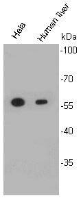
Fig1: Western blot analysis on Hela cell and human liver lysates using anti- ALDH2 rabbit polyclonal antibodies.Application
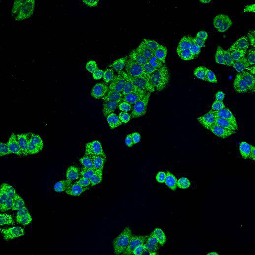
Fig2: ICC staining ALDH2 in Hela cells (green). Cells were fixed in paraformaldehyde, permeabilised with 0.25% Triton X100/PBS and counterstained with DAPI in order to highlight the nucleus (blue).Application
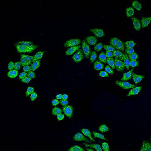
Fig3: ICC staining ALDH2 in HepG2 cells (green). Cells were fixed in paraformaldehyde, permeabilised with 0.25% Triton X100/PBS and counterstained with DAPI in order to highlight the nucleus (blue).| Product Name | ALDH2 |
|---|---|
| Antibody Type | Primary Antibodies |
| Antigen Alias | Acetaldehyde dehydrogenase 2 antibody Aldehyde dehydrogenase 2 family (mitochondrial) antibody Aldehyde dehydrogenase 2 family antibody Aldehyde dehydrogenase mitochondrial antibody Aldehyde dehydrogenase, mitochondrial antibody ALDH 2 antibody ALDH class 2 antibody ALDH E2 antibody ALDH-E2 antibody Aldh2 antibody ALDH2_HUMAN antibody ALDHI antibody ALDM antibody Liver mitochondrial ALDH antibody MGC1806 antibody Mitochondrial aldehyde dehydrogenase 2 antibody MS767 antibody Nucleus encoded mitochondrial aldehyde dehydrogenase 2 antibody |
| Product description | Aldehyde dehydrogenase is a?polymorphic?enzyme ?responsible for the?oxidation?of?aldehydes?tocarboxylic acids, which leave the liver and are metabolized by the body’s muscle and heart. The active site of the aldehyde dehydrogenase enzyme is largely conserved throughout the different classes of the enzyme and, although the number of amino acids present in a subunit can change, the overall function of the site changes little. ALDH2 plays a crucial role in maintaining low blood levels of acetaldehyde during alcohol oxidation. In this pathway, the intermediate structures can be toxic, and health problems arise when those intermediates cannot be cleared.?When high levels of acetaldehyde occur in the blood, facial flushing, light headedness, palpitations, nausea, and general “hangover” symptoms occur. These symptoms are indicative of a disease known as the?Alcohol flush reaction, also known as “Asian Flush” or “Oriental Flushing Syndrome”. |
| Immunogen | peptide |
| Clonality | Polyclonal |
|---|---|
| Isotype | IgG |
| Host Species | Rabbit |
| Tested Applications | WBICC |
| WB:1:1,000 ICC:1:200 |
|
| Species Reactivity | Human |
| Concentration | 1mg/ml |
| Alternative Names | Acetaldehyde dehydrogenase 2 antibody
Aldehyde dehydrogenase 2 family (mitochondrial) antibody
Aldehyde dehydrogenase 2 family antibody
Aldehyde dehydrogenase mitochondrial antibody
Aldehyde dehydrogenase mitochondrial antibody ALDH 2 antibody ALDH class 2 antibody ALDH E2 antibody ALDH-E2 antibody Aldh2 antibody ALDH2_HUMAN antibody ALDHI antibody ALDM antibody Liver mitochondrial ALDH antibody MGC1806 antibody Mitochondrial aldehyde dehydrogenase 2 antibody MS767 antibody Nucleus encoded mitochondrial aldehyde dehydrogenase 2 antibody |
|---|---|
| Molecular Weight(MW) | 56 kDa |
| Cellular Localization | Mitochondrion matrix |
| SwissProt ID | P05091 |
|---|
Application
Fig1: Western blot analysis on Hela cell and human liver lysates using anti- ALDH2 rabbit polyclonal antibodies.
Application
Fig2: ICC staining ALDH2 in Hela cells (green). Cells were fixed in paraformaldehyde, permeabilised with 0.25% Triton X100/PBS and counterstained with DAPI in order to highlight the nucleus (blue).
Application
Fig3: ICC staining ALDH2 in HepG2 cells (green). Cells were fixed in paraformaldehyde, permeabilised with 0.25% Triton X100/PBS and counterstained with DAPI in order to highlight the nucleus (blue).| Positive Control | Hela, HepG2 , human liver |
|---|---|
| Application Notes | WB:1:1,000 ICC:1:200 |
| Form | Liquid |
|---|---|
| Storage Instructions | Store at +4℃ after thawing. Aliquot store at -20℃ or -80℃. Avoid repeated freeze / thaw cycles. |
| Storage Buffer | 1*TBS (pH7.4), 1%BSA, 40%Glycerol. Preservative: 0.05% Sodium Azide. |


