Application
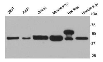
Fig1: Western blot analysis on different cell and tissue lysates using anti-AKR7A2 rabbit polyclonal antibodies.Application
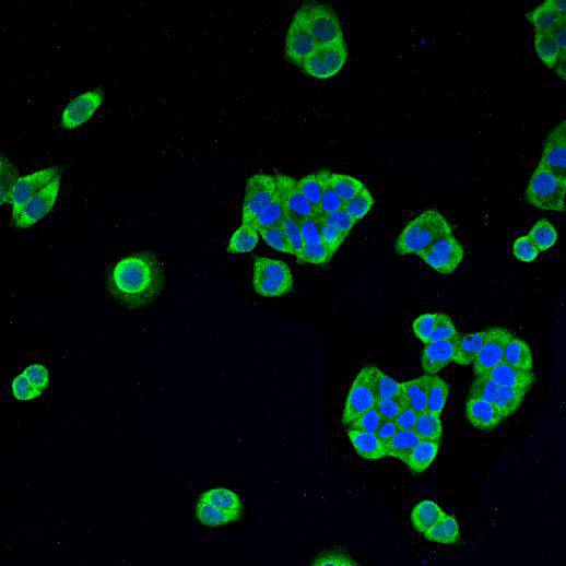
Fig2: ICC staining AKR7A2 in Hela cells (green). Cells were fixed in paraformaldehyde, permeabilised with 0.25% Triton X100/PBS and counterstained with DAPI in order to highlight the nucleus (blue).| Product Name | AKR7A2 |
|---|---|
| Antibody Type | Primary Antibodies |
| Antigen Alias | AFAR antibody AFAR1 antibody AFB1 aldehyde reductase 1 antibody AFB1 AR1 antibody AFB1-AR 1 antibody AFB1AR1 antibody Aflatoxin aldehyde reductase antibody Aflatoxin B1 aldehyde reductase member 2 antibody Aflatoxin beta1 aldehyde reductase antibody Aiar antibody AKR7 antibody Akr7a2 antibody Aldo keto reductase family 7 antibody Aldo keto reductase family 7 member A2 aflatoxin aldehyde reductase antibody Aldo keto reductase family 7 member A2 antibody Aldo keto reductase family 7, member A2 (aflatoxin aldehyde reductase) antibody Aldoketoreductase 7 antibody ARK72_HUMAN antibody SSA reductase antibody Succinic semialdehyde reductase antibody |
| Product description | Aflatoxin B1 aldehyde reductase member 2 is an enzyme that in humans is encoded by the AKR7A2 gene. It catalyzes the NADPH-dependent reduction of succinic semialdehyde to gamma-hydroxybutyrate and may have an important role in producing the neuromodulator gamma-hydroxybutyrate (GHB). AKR7A2 has broad substrate specificity and NADPH-dependent aldehyde reductase activity towards 2-carboxybenzaldehyde, 2-nitrobenzaldehyde and pyridine-2-aldehyde (in vitro). AKR7A2 can reduce 1,2-naphthoquinone and 9,10-phenanthrenequinone (in vitro) and reduce the dialdehyde protein-binding form of aflatoxin B1 (AFB1) to the non-binding AFB1 dialcohol. It may be involved in protection of liver against the toxic and carcinogenic effects of AFB1, a potent hepatocarcinogen. |
| Immunogen | peptide |
| Clonality | Polyclonal |
|---|---|
| Isotype | IgG |
| Host Species | Rabbit |
| Tested Applications | WBICC |
| WB:1:1000-1:2000 ICC:1:200 |
|
| Species Reactivity | HumanMouseRat |
| Concentration | 1mg/ml |
| Alternative Names | AFAR antibody
AFAR1 antibody
AFB1 aldehyde reductase 1 antibody
AFB1 AR1 antibody
AFB1-AR 1 antibody
AFB1AR1 antibody
Aflatoxin aldehyde reductase antibody
Aflatoxin B1 aldehyde reductase member 2 antibody
Aflatoxin beta1 aldehyde reductase antibody
Aiar antibody
AKR7 antibody
Akr7a2 antibody
Aldo keto reductase family 7 antibody
Aldo keto reductase family 7 member A2 aflatoxin aldehyde reductase antibody
Aldo keto reductase family 7 member A2 antibody
Aldo keto reductase family 7 member A2 (aflatoxin aldehyde reductase) antibody Aldoketoreductase 7 antibody ARK72_HUMAN antibody SSA reductase antibody Succinic semialdehyde reductase antibody |
|---|---|
| Molecular Weight(MW) | 40 kDa |
| Cellular Localization | Golgi apparatus, cytoplasm |
| SwissProt ID | O43488 |
|---|
Application
Fig1: Western blot analysis on different cell and tissue lysates using anti-AKR7A2 rabbit polyclonal antibodies.
Application
Fig2: ICC staining AKR7A2 in Hela cells (green). Cells were fixed in paraformaldehyde, permeabilised with 0.25% Triton X100/PBS and counterstained with DAPI in order to highlight the nucleus (blue).| Positive Control | 293T, A431, Jurkat, mouse liver, rat liver, human liver, Hela |
|---|---|
| Application Notes | WB:1:1000-1:2000 ICC:1:200 |
| Form | Liquid |
|---|---|
| Storage Instructions | Store at +4℃ after thawing. Aliquot store at -20℃ or -80℃. Avoid repeated freeze / thaw cycles. |
| Storage Buffer | 1*TBS (pH7.4), 1%BSA, 40%Glycerol. Preservative: 0.05% Sodium Azide. |


