Application
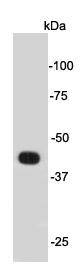
Fig1: Western blot analysis on mouse brain cell lysates using anti- Psg16 rabbit polyclonal antibodies.Application
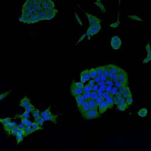
Fig2: ICC staining Psg16 in mouse embryonic stem cells D3 (green). Cells were fixed in paraformaldehyde, permeabilised with 0.25% Triton X100/PBS and counterstained with DAPI in order to highlight the nucleus (blue).| Product Name | Psg16 |
|---|---|
| Antibody Type | Primary Antibodies |
| Antigen Alias | Pregnancy specific glycoprotein 16, Pregnancy-specific glycoprotein 16 |
| Product description | The murine pregnancy-specific glycoprotein-16 (PSG16; previously brain carcinoembryonic antigen/bCEA) is expressed in the brain, especially in neurons, and has previously been reported to be an MHV receptor. The murine pregnancy-specific glycoproteins (PSGs), which mediate placentation and modulate interactions between the fetus and the maternal immune system, resemble the related carcinoembryonic antigen (CEA) proteins in their extracellular Ig-like domains but are secreted rather than membrane-anchored. |
| Immunogen | peptide |
| Clonality | Polyclonal |
|---|---|
| Isotype | IgG |
| Host Species | Rabbit |
| Tested Applications | WBICCFC |
| WB:1:1000 ICC:1:100-1:200 |
|
| Species Reactivity | Mouse |
| Concentration | 1mg/ml |
| Alternative Names | Pregnancy specific glycoprotein 16 Pregnancy-specific glycoprotein 16 |
|---|---|
| Molecular Weight(MW) | 45 kDa |
| Cellular Localization | Cell membrane, Cell projection |
| SwissProt ID | Q8K0U8 |
|---|
Application
Fig1: Western blot analysis on mouse brain cell lysates using anti- Psg16 rabbit polyclonal antibodies.
Application
Fig2: ICC staining Psg16 in mouse embryonic stem cells D3 (green). Cells were fixed in paraformaldehyde, permeabilised with 0.25% Triton X100/PBS and counterstained with DAPI in order to highlight the nucleus (blue).| Positive Control | Mouse brain, D3 |
|---|---|
| Application Notes | WB:1:1000 ICC:1:100-1:200 |
| Form | Liquid |
|---|---|
| Storage Instructions | Store at +4℃ after thawing. Aliquot store at -20℃ or -80℃. Avoid repeated freeze / thaw cycles. |
| Storage Buffer | 1*TBS (pH7.4), 1%BSA, 40%Glycerol. Preservative: 0.05% Sodium Azide. |


