Application
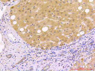
Fig1: Immunohistochemical analysis of paraffin-embedded human liver carcinoma tissue using anti- Beclin 1 rabbit polyclonal antibody.Application
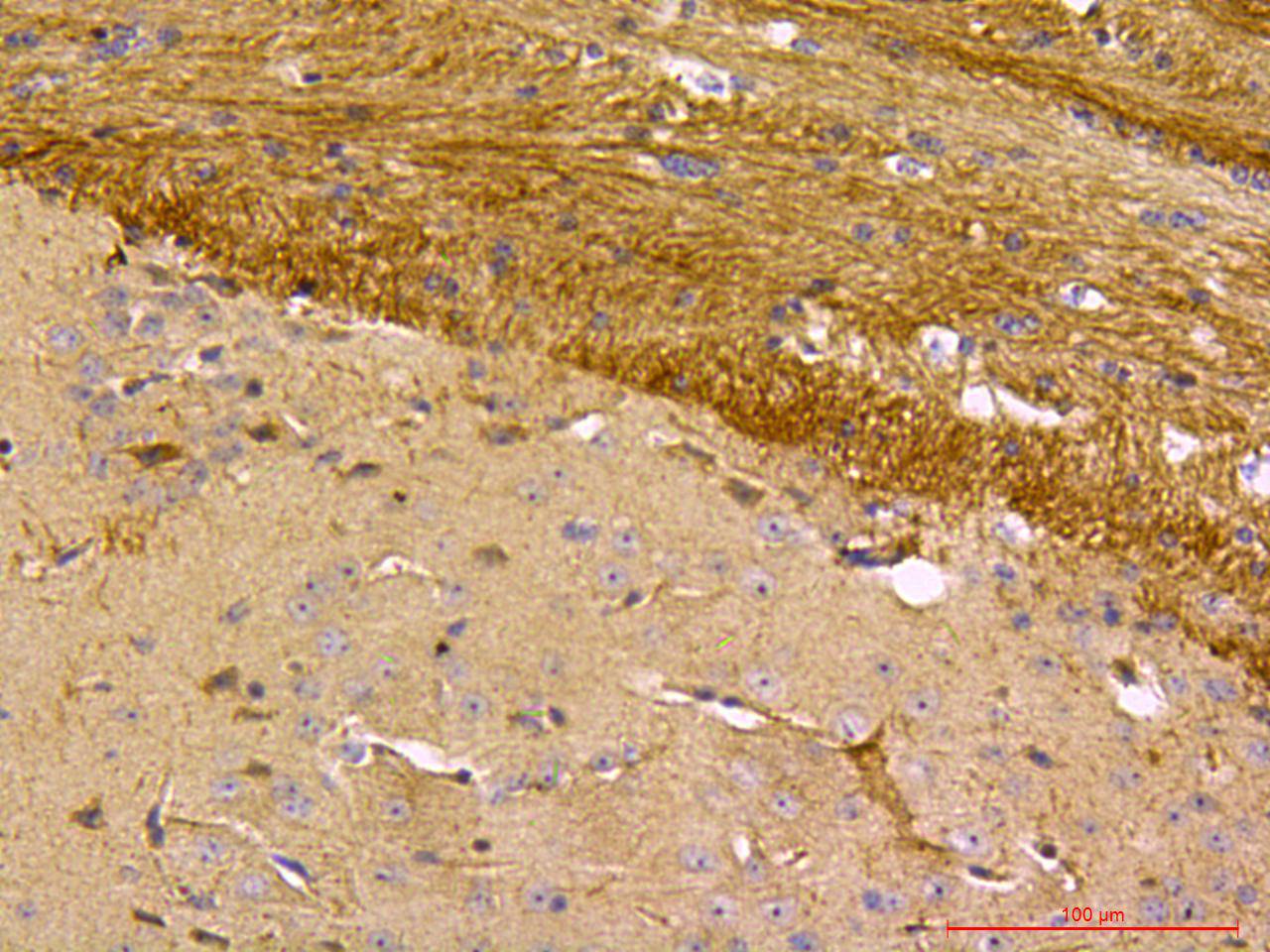
Fig2: Immunohistochemical analysis of paraffin-embedded mouse brain tissue using anti- Beclin 1 rabbit polyclonal antibody.Application
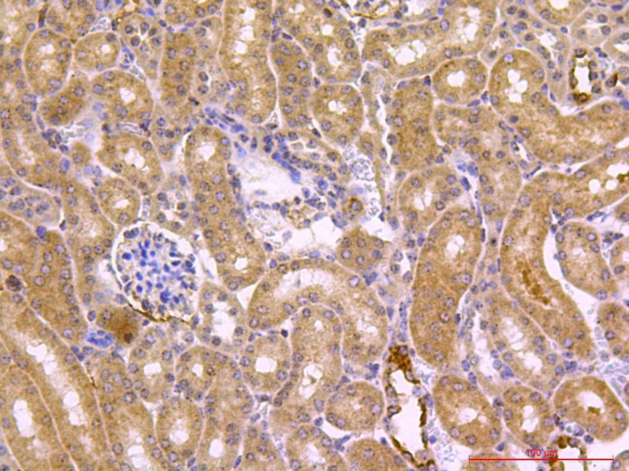
Fig3: Immunohistochemical analysis of paraffin-embedded mouse kidney tissue using anti- Beclin 1 rabbit polyclonal antibody.Application
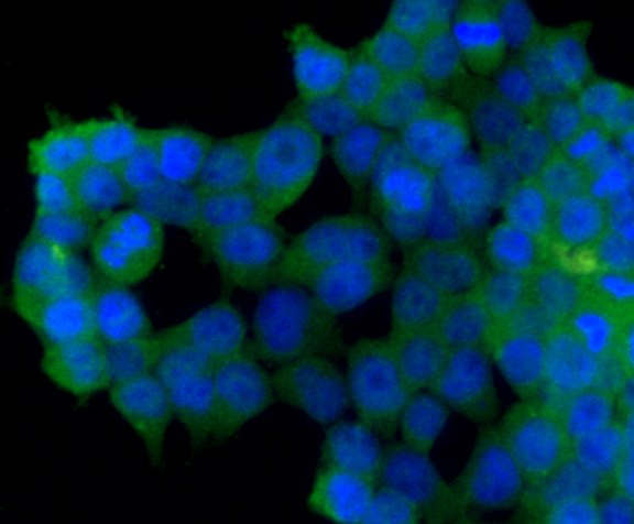
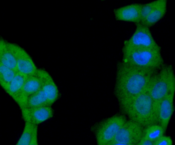
Fig4: ICC staining Beclin 1 in Ags cells (green). The nuclear counter stain is DAPI (blue). Cells were fixed in paraformaldehyde, permeabilised with 0.25% Triton X100/PBS.Application
Fig5: ICC staining Beclin 1 in Hela cells (green). The nuclear counter stain is DAPI (blue). Cells were fixed in paraformaldehyde, permeabilised with 0.25% Triton X100/PBS.| Product Name | Beclin 1 |
|---|---|
| Antibody Type | Primary Antibodies |
| Antigen Alias | APG6 antibody ATG 6 antibody ATG6 antibody ATG6 autophagy related 6 homolog antibody Bcl-2-interacting protein beclin antibody Beclin 1 (coiled coil moesin like BCL2 interacting protein) antibody Beclin 1 autophagy related antibody Beclin-1 antibody Beclin1 antibody BECN 1 antibody Becn1 antibody BECN1_HUMAN antibody Coiled coil myosin like BCL2 interacting protein antibody Coiled-coil myosin-like BCL2-interacting protein antibody GT197 antibody Protein GT197 antibody VPS 30 antibody VPS30 antibody |
| Product description | Plays a central role in autophagy. Acts as core subunit of the PI3K complex that mediates formation of phosphatidylinositol 3-phosphate; different complex forms are believed to play a role in multiple membrane trafficking pathways: PI3KC3-C1 is involved in initiation of autophagosomes and PI3KC3-C2 in maturation of autophagosomes and endocytosis. Involved in regulation of degradative endocytic trafficking and required for the abcission step in cytokinesis, probably in the context of PI3KC3-C2. Essential for the formation of PI3KC3-C2 but not PI3KC3-C1 PI3K complex forms. Involved in endocytosis. Protects against infection by a neurovirulent strain of Sindbis virus. May play a role in antiviral host defense. |
| Immunogen | Synthetic peptide (KLH-coupled) within human Beclin 1 aa 380-450. |
| Clonality | Polyclonal |
|---|---|
| Isotype | IgG |
| Host Species | Rabbit |
| Tested Applications | ICCIHC |
| IHC:1:200 ICC:1:50-1:100 |
|
| Species Reactivity | HumanMouseRat |
| Concentration | 1mg/ml |
| Alternative Names | APG6 antibody ATG 6 antibody ATG6 antibody ATG6 autophagy related 6 homolog antibody Bcl-2-interacting protein beclin antibody Beclin 1 (coiled coil moesin like BCL2 interacting protein) antibody Beclin 1 autophagy related antibody Beclin-1 antibody Beclin1 antibody BECN 1 antibody Becn1 antibody BECN1_HUMAN antibody Coiled coil myosin like BCL2 interacting protein antibody Coiled-coil myosin-like BCL2-interacting protein antibody GT197 antibody Protein GT197 antibody VPS 30 antibody VPS30 antibody |
|---|---|
| Molecular Weight(MW) | 52kDa |
Application
Fig1: Immunohistochemical analysis of paraffin-embedded human liver carcinoma tissue using anti- Beclin 1 rabbit polyclonal antibody.
Application
Fig2: Immunohistochemical analysis of paraffin-embedded mouse brain tissue using anti- Beclin 1 rabbit polyclonal antibody.
Application
Fig3: Immunohistochemical analysis of paraffin-embedded mouse kidney tissue using anti- Beclin 1 rabbit polyclonal antibody.
Application
Fig4: ICC staining Beclin 1 in Ags cells (green). The nuclear counter stain is DAPI (blue). Cells were fixed in paraformaldehyde, permeabilised with 0.25% Triton X100/PBS.
Application
Fig5: ICC staining Beclin 1 in Hela cells (green). The nuclear counter stain is DAPI (blue). Cells were fixed in paraformaldehyde, permeabilised with 0.25% Triton X100/PBS.| Positive Control | Human liver carcinoma tissue, mouse brain, mouse kidney, Ags, Hela. |
|---|---|
| Application Notes | IHC:1:200 ICC:1:50-1:100 |
| Form | Liquid |
|---|---|
| Storage Instructions | Store at +4℃ after thawing. Aliquot store at -20℃. Avoid repeated freeze / thaw cycles. |
| Storage Buffer | 1*TBS (pH7.4), 1%BSA, 40%Glycerol. Preservative: 0.05% Sodium Azide. |


