Application
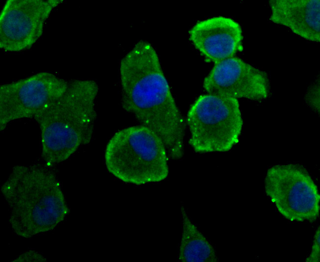
Fig1: ICC staining Rem2 in A549 cells (green). The nuclear counter stain is DAPI (blue). Cells were fixed in paraformaldehyde, permeabilised with 0.25% Triton X100/PBS.Application
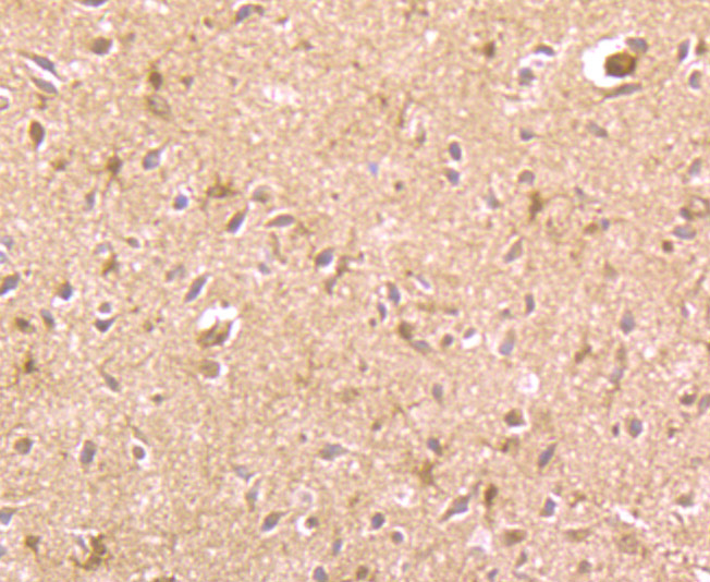
Fig2: Immunohistochemical analysis of paraffin-embedded rat brain tissue using anti-Rem2 antibody. Counter stained with hematoxylin.Application
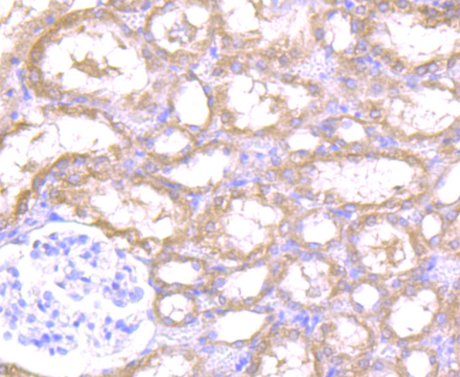
Fig3: Immunohistochemical analysis of paraffin-embedded rat kidney tissue using anti-Rem2 antibody. Counter stained with hematoxylin.Application
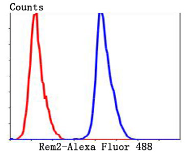
Fig4: Flow cytometric analysis of HepG2 cells with Rem2 antibody at 1/100 dilution (blue) compared with an unlabelled control (cells without incubation with primary antibody; red). Alexa Fluor® 488-conjugated goat anti-rabbit IgG was used as the secondary antibody.| Product Name | Rem2 |
|---|---|
| Antibody Type | Primary Antibodies |
| Antigen Alias | cb111 antibody chunp6884 antibody etID309896.20 antibody R2 antibody reductase M2 polypeptide variant 1 antibody reductase M2 polypeptide variant 2 antibody reductase M2 polypeptide variant 3a antibody reductase M2 polypeptide variant 3b antibody reductase M2 polypeptide variant 3c antibody reductase M2 polypeptide variant 3d antibody Ribonucleoside-diphosphate reductase subunit M2 antibody Ribonucleotide reductase M2 antibody Ribonucleotide reductase M2 polypeptide antibody Ribonucleotide reductase M2 subunit antibody ribonucleotide reductase protein r2 class I antibody Ribonucleotide reductase regulatory subunit M2 antibody Ribonucleotide reductase small chain antibody Ribonucleotide reductase small subunit antibody Ribonucleotide reductase, R2 subunit antibody RIR2_HUMAN antibody RR2 antibody RR2M antibody RRM2 antibody |
| Product description | REM2 (RAS (RAD and GEM)-like GTP binding 2) is a 340 amino acid cell membrane protein that functions as a GTPase and belongs to the RGK family as well as the small GTPase superfamily. A suppressor of the p53 pathway, REM2 also mediates the fibroblastic growth factor 2 (FGF-2) signaling pathway and is involved in maintaining the proliferation of human embryonic stem cells. Expressed in kidney and brain, REM2 is encoded by a gene that maps to human chromosome 14q11.2. Chromosome 14 encodes the presinilin 1 (PSEN1) gene, which is one of the three key genes associated with the development of Alzheimer's disease (AD). The SERPINA1 gene is also located on chromosome 14 and, when defective, leads to the genetic disorder α1-antitrypsin deficiency, which is characterized by severe lung complications and liver dysfunction. |
| Immunogen | Recombinant protein |
| Clonality | Polyclonal |
|---|---|
| Isotype | IgG |
| Host Species | Rabbit |
| Tested Applications | IHCFC |
| ICC:1:50-1:200 IHC:1:50-1:200 FC:1:50-1:100 WB:1:500 |
|
| Species Reactivity | Human |
| Concentration | 1mg/ml |
| Alternative Names | cb111 antibody
chunp6884 antibody
etID309896.20 antibody
R2 antibody
reductase M2 polypeptide variant 1 antibody
reductase M2 polypeptide variant 2 antibody
reductase M2 polypeptide variant 3a antibody
reductase M2 polypeptide variant 3b antibody
reductase M2 polypeptide variant 3c antibody
reductase M2 polypeptide variant 3d antibody
Ribonucleoside-diphosphate reductase subunit M2 antibody
Ribonucleotide reductase M2 antibody
Ribonucleotide reductase M2 polypeptide antibody
Ribonucleotide reductase M2 subunit antibody
ribonucleotide reductase protein r2 class I antibody
Ribonucleotide reductase regulatory subunit M2 antibody
Ribonucleotide reductase small chain antibody
Ribonucleotide reductase small subunit antibody
Ribonucleotide reductase R2 subunit antibody RIR2_HUMAN antibody RR2 antibody RR2M antibody RRM2 antibody |
|---|---|
| Molecular Weight(MW) | 37 kDa |
| Cellular Localization | Cell membrane. |
| SwissProt ID | Q8IYK8 |
|---|
Application
Fig1: ICC staining Rem2 in A549 cells (green). The nuclear counter stain is DAPI (blue). Cells were fixed in paraformaldehyde, permeabilised with 0.25% Triton X100/PBS.
Application
Fig2: Immunohistochemical analysis of paraffin-embedded rat brain tissue using anti-Rem2 antibody. Counter stained with hematoxylin.
Application
Fig3: Immunohistochemical analysis of paraffin-embedded rat kidney tissue using anti-Rem2 antibody. Counter stained with hematoxylin.
Application
Fig4: Flow cytometric analysis of HepG2 cells with Rem2 antibody at 1/100 dilution (blue) compared with an unlabelled control (cells without incubation with primary antibody; red). Alexa Fluor® 488-conjugated goat anti-rabbit IgG was used as the secondary antibody.| Positive Control | A549, HepG2, rat brain tissue, rat kidney tissue. |
|---|---|
| Application Notes | ICC:1:50-1:200 IHC:1:50-1:200 FC:1:50-1:100 WB:1:500 |
| Form | Liquid |
|---|---|
| Storage Instructions | Store at +4℃ after thawing. Aliquot store at -20℃ or -80℃. Avoid repeated freeze / thaw cycles. |
| Storage Buffer | 1*TBS (pH7.4), 1%BSA, 40%Glycerol. Preservative: 0.05% Sodium Azide. |


