Application
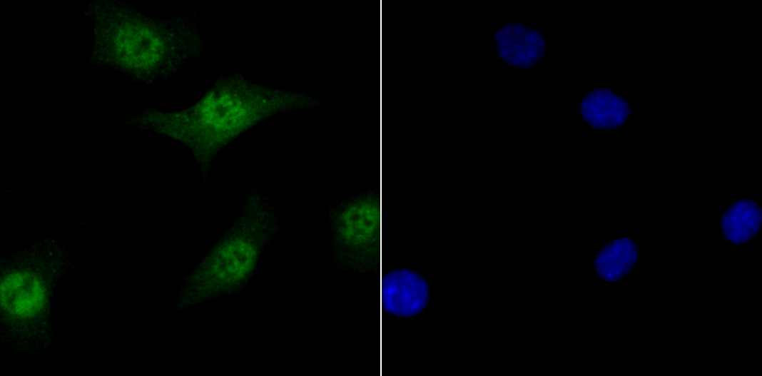
Fig1: Immunocytochemical staining of SH-SY-5Y cells using anti-TET1 rabbit polyclonal antibody.Application
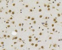
Fig2: Immunohistochemical analysis of paraffin- embedded mouse brain tissue using anti-TET1 rabbit polyclonal antibody.Application
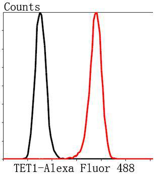
Fig3: Flow cytometric analysis of SH-SY-5Y cells with TET1 antibody at 1/50 dilution (red) compared with an unlabelled control (cells without incubation with primary antibody; black). Alexa Fluor 488-conjugated Goat anti rabbit IgG was used as the secondary antibody.| Product Name | Tet1 |
|---|---|
| Antibody Type | Primary Antibodies |
| Antigen Alias | bA119F7.1 antibody CXXC 6 antibody CXXC finger 6 antibody CXXC type zinc finger protein 6 antibody CXXC zinc finger 6 antibody CXXC-type zinc finger protein 6 antibody CXXC6 antibody KIAA1676 antibody LCX antibody Leukemia associated protein with a CXXC domain antibody Leukemia-associated protein with a CXXC domain antibody Methylcytosine dioxygenase TET1 antibody Ten eleven translocation 1 antibody Ten eleven translocation 1 gene protein antibody Ten eleven translocation 1 gene protein homolog antibody Ten-eleven translocation 1 gene protein antibody Tet 1 antibody Tet methylcytosine dioxygenase 1 antibody Tet oncogene 1 antibody TET1 antibody TET1_HUMAN antibody |
| Product description | TET1 (tet oncogene 1), also known as LCX or CXXC6, is a 2,136 amino acid protein that localizes to the nucleus and contains one CXXC-type zinc finger. Expressed in adult ovary, thymus and skeletal muscle and also present in fetal lung, heart and brain, TET1 is thought to play a role in the development of fetal organs and may also be involvement in the pathoegenesis and metastasis of acute myeloid leukemia (AML). The gene encoding TET1 maps to human chromosome 10, which houses over 1,200 genes and comprises nearly 4.5% of the human genome. Defects in some of the genes that map to chromosome 10 are associated with Charcot-Marie Tooth disease, Jackson-Weiss syndrome, Usher syndrome, nonsyndromatic deafness, Wolman’s syndrome, Cowden syndrome, multiple endocrine neoplasia type 2 and porphyria. |
| Immunogen | Synthetic peptide (KLH-coupled) within human TET1 820-900 aa. |
| Clonality | Polyclonal |
|---|---|
| Isotype | IgG |
| Host Species | Rabbit |
| Tested Applications | IHCFCICC |
| ICC:1:50-1:200 IHC:1:50-1:200 FC:1:50-1:100 |
|
| Species Reactivity | HumanMouseRat |
| Concentration | 1mg/ml |
| Alternative Names | bA119F7.1 antibody CXXC 6 antibody CXXC finger 6 antibody CXXC type zinc finger protein 6 antibody CXXC zinc finger 6 antibody CXXC-type zinc finger protein 6 antibody CXXC6 antibody KIAA1676 antibody LCX antibody Leukemia associated protein with a CXXC domain antibody Leukemia-associated protein with a CXXC domain antibody Methylcytosine dioxygenase TET1 antibody Ten eleven translocation 1 antibody Ten eleven translocation 1 gene protein antibody Ten eleven translocation 1 gene protein homolog antibody Ten-eleven translocation 1 gene protein antibody Tet 1 antibody Tet methylcytosine dioxygenase 1 antibody Tet oncogene 1 antibody TET1 antibody TET1_HUMAN antibody |
|---|---|
| Molecular Weight(MW) | 235kDa |
| Cellular Localization | Nucleus |
Application
Fig1: Immunocytochemical staining of SH-SY-5Y cells using anti-TET1 rabbit polyclonal antibody.
Application
Fig2: Immunohistochemical analysis of paraffin- embedded mouse brain tissue using anti-TET1 rabbit polyclonal antibody.
Application
Fig3: Flow cytometric analysis of SH-SY-5Y cells with TET1 antibody at 1/50 dilution (red) compared with an unlabelled control (cells without incubation with primary antibody; black). Alexa Fluor 488-conjugated Goat anti rabbit IgG was used as the secondary antibody.| Positive Control | SH-SY-5Y, mouse brain tissue. |
|---|---|
| Application Notes | ICC:1:50-1:200 IHC:1:50-1:200 FC:1:50-1:100 |
| Form | Liquid |
|---|---|
| Storage Instructions | Store at +4℃ after thawing. Aliquot store at -20℃. Avoid repeated freeze / thaw cycles. |
| Storage Buffer | 1*TBS (pH7.4), 1%BSA, 40%Glycerol. Preservative: 0.05% Sodium Azide. |


