Application
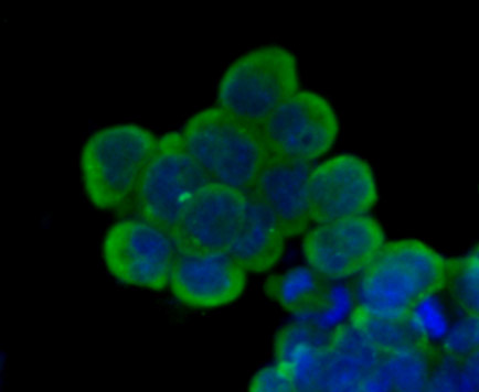
Fig1: ICC staining Nestin in F9 cells (green). The nuclear counter stain is DAPI (blue). Cells were fixed in paraformaldehyde, permeabilised with 0.25% Triton X100/PBS.Application
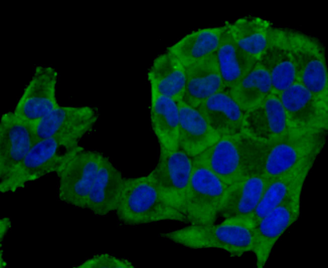
Fig2: ICC staining Nestin in Hela cells (green). The nuclear counter stain is DAPI (blue). Cells were fixed in paraformaldehyde, permeabilised with 0.25% Triton X100/PBS.Application
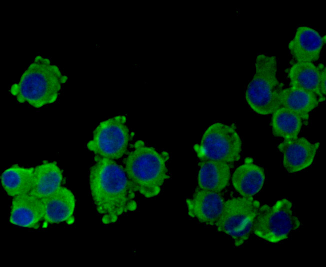
Fig3: ICC staining Nestin in N2A cells (green). The nuclear counter stain is DAPI (blue). Cells were fixed in paraformaldehyde, permeabilised with 0.25% Triton X100/PBS.Application
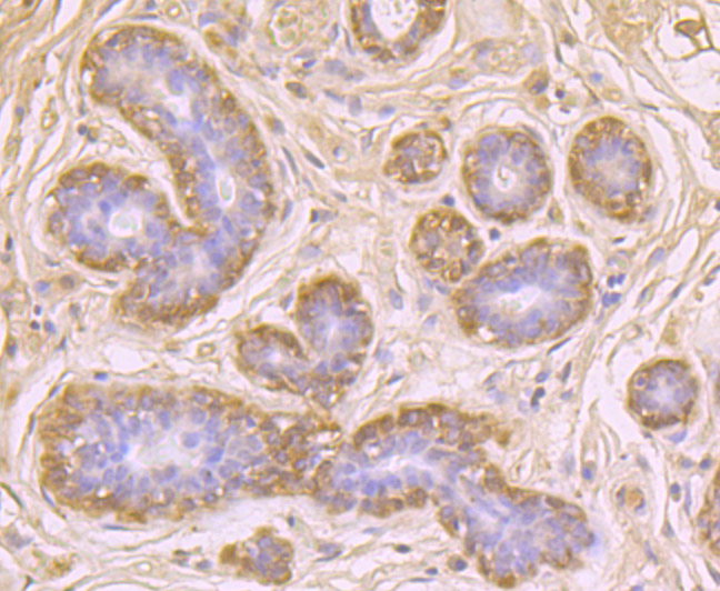
Fig4: Immunohistochemical analysis of paraffin-embedded human breast tissue using anti-Nestin antibody. Counter stained with hematoxylin.Application
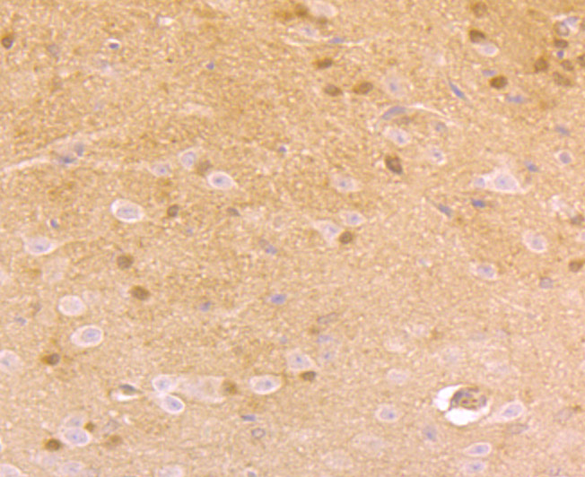
Fig5: Immunohistochemical analysis of paraffin-embedded mouse brain tissue using anti-Nestin antibody. Counter stained with hematoxylin.Application
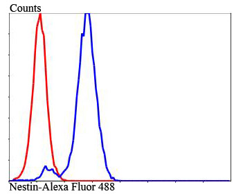
Fig6: Flow cytometric analysis of N2A cells with Nestin antibody at 1/100 dilution (blue) compared with an unlabelled control (cells without incubation with primary antibody; red). Alexa Fluor® 488-conjugated goat anti-rabbit IgG was used as the secondary antibody.| Product Name | Nestin |
|---|---|
| Antibody Type | Primary Antibodies |
| Antigen Alias | ESTM 46 antibody FLJ 21841 antibody FLJ21841 antibody Intermediate filament protein antibody Nbla00170 antibody nes antibody NEST_HUMAN antibody Nestin antibody |
| Product description | Nestin is a major intermediate filament (IF) protein of embryonic central nervous system progenitor cells. It is also a component of the dynamic IF network during muscle development, where it polymerizes with Desmin and Vimentin. Nestin co-assembles with Vimentin or a-internexin and forms heterodimer coiled-coil molecules which then further assemble into 10 nml IFs. Deletion of the IF consensus rod domain in nestin alters nestin localization in CNS precursor cells and radial glial cells in vivo. Nestin is a marker for neuroepithelial stem cells, glioma cells and tumor endothelial cells during rapid growth. During axon elongation of differen-tiation neurons, nestin localizes to the growth cones and may play a role in growth cone guidance. In the rat adrenal gland, nestin is expressed by the zona fasciculata and the zona reticularis. Nestin is also expressed by dermatomal cells and by myoblasts during the earliest stages of myogenesis. |
| Immunogen | Peptide |
| Clonality | Polyclonal |
|---|---|
| Isotype | IgG |
| Host Species | Rabbit |
| Tested Applications | ICC |
| ICC:1:200~1:500 Notes:Optimal dilutions/concentrations should be determined by the researcher. |
|
| Species Reactivity | HumanMouse |
| Concentration | 1mg/ml |
| Alternative Names | ESTM 46 antibody FLJ 21841 antibody FLJ21841 antibody Intermediate filament protein antibody Nbla00170 antibody nes antibody NEST_HUMAN antibody Nestin antibody |
|---|---|
| Molecular Weight(MW) | 177 kDa(Predicted band size) |
| Cellular Localization | Cytoskeleton, extracellular region or secreted. |
| SwissProt ID | Q6P5H2 |
|---|
Application
Fig1: ICC staining Nestin in F9 cells (green). The nuclear counter stain is DAPI (blue). Cells were fixed in paraformaldehyde, permeabilised with 0.25% Triton X100/PBS.
Application
Fig2: ICC staining Nestin in Hela cells (green). The nuclear counter stain is DAPI (blue). Cells were fixed in paraformaldehyde, permeabilised with 0.25% Triton X100/PBS.
Application
Fig3: ICC staining Nestin in N2A cells (green). The nuclear counter stain is DAPI (blue). Cells were fixed in paraformaldehyde, permeabilised with 0.25% Triton X100/PBS.
Application
Fig4: Immunohistochemical analysis of paraffin-embedded human breast tissue using anti-Nestin antibody. Counter stained with hematoxylin.
Application
Fig5: Immunohistochemical analysis of paraffin-embedded mouse brain tissue using anti-Nestin antibody. Counter stained with hematoxylin.
Application
Fig6: Flow cytometric analysis of N2A cells with Nestin antibody at 1/100 dilution (blue) compared with an unlabelled control (cells without incubation with primary antibody; red). Alexa Fluor® 488-conjugated goat anti-rabbit IgG was used as the secondary antibody.| Positive Control | F9, Hela, N2A, human breast tissue, mouse brain tissue. |
|---|---|
| Application Notes | ICC:1:200~1:500 Notes:Optimal dilutions/concentrations should be determined by the researcher. |
| Form | Liquid |
|---|---|
| Storage Instructions | Store at +4℃ after thawing. Aliquot store at -20℃ or -80℃. Avoid repeated freeze / thaw cycles. |
| Storage Buffer | 1*TBS (pH7.4), 1%BSA, 40%Glycerol. Preservative: 0.05% Sodium Azide. |


