Application
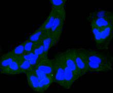
Fig1: Immunocytochemical staining of Hela cells using anti-M6PR rabbit polyclonal antibody.Application
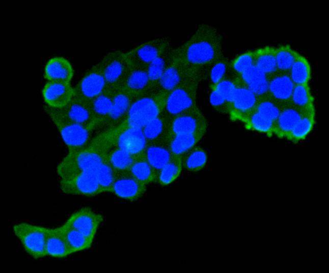
Fig2: Immunocytochemical staining of SW480 cells using anti-M6PR rabbit polyclonal antibody.Application
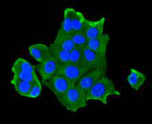
Fig3: Immunocytochemical staining of MCF-7 cells using anti-M6PR rabbit polyclonal antibody.Application
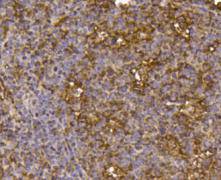
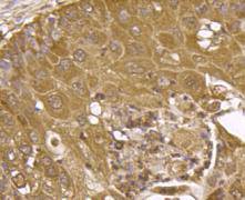
Fig4: Immunohistochemical analysis of paraffin- embedded human tonsil tissue using anti-M6PR rabbit polyclonal antibody.Application
Fig5: Immunohistochemical analysis of paraffin- embedded human lung carcinoma tissue using anti-M6PR rabbit polyclonal antibody.Application
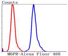
Fig6: Flow cytometric analysis of Hela cells with M6PR antibody at 1/50 dilution (blue) compared with an unlabelled control (cells without incubation with primary antibody; red). Alexa Fluor 488-conjugated Goat anti mouse IgG was used as the secondary antibody.| Product Name | IGF2R/M6PR |
|---|---|
| Antibody Type | Primary Antibodies |
| Antigen Alias | 300 kDa mannose 6 phosphate receptor antibody 300 kDa mannose 6-phosphate receptor antibody Cation independent mannose 6 phosphate receptor antibody Cation-independent mannose-6-phosphate receptor antibody CD222 antibody CD222 antigen antibody CI Man 6 P receptor antibody CI Man-6-P receptor antibody CI MPR antibody CI-M6PR antibody CI-MPR antibody CIMPR antibody IGF 2 receptor antibody IGF 2R antibody IGF II receptor antibody IGF-II receptor antibody IGF2 receptor antibody Igf2r antibody Insulin like growth factor 2 receptor antibody Insulin like growth factor II receptor antibody Insulin-like growth factor 2 receptor antibody Insulin-like growth factor II receptor antibody M6P R antibody M6P/IGF2 receptor antibody M6P/IGF2R antibody M6PR antibody mannose 6 phosphate receptor antibody mannose 6 phosphate receptor, cation independent antibody MPR 300 antibody MPR300 antibody MPRI antibody MPRI_HUMAN antibody |
| Product description | The mannose 6-phosphate/insulin-like growth factor II receptor, IGF-IIR (also designated M6P/IGF2R), is a ubiquitously expressed integral glycoprotein. By binding glycoproteins through two of its extracytoplasmic domains, IGF-IIR mediates the activation of TGF∫1 (a growth inhibitor), the degradation of IGF-II and the transport of lysosomal enzymes. Subsequently, IGF-IIR can form oligomeric complexes, which increase the affinity of IGF-IIR for lysosomal enzymes. Unlike IGF-IR, IGF-IIR does not potentiate the signaling of IGF-I or IGF-II, which have mitogenic, cell survival and insulin-like effects. Therefore, IGF-IIR is characterized as a tumor suppressor. Furthermore, the IGF-IIR gene is located on chromosome 6q26, which is commonly mutated or deleted in several human cancers. |
| Immunogen | recombinant protein |
| Clonality | Polyclonal |
|---|---|
| Isotype | IgG |
| Host Species | Rabbit |
| Tested Applications | ICCIHCFC |
| ICC:1:50-1:200 IHC:1:50-1:200 FC:1:50-1:100 WB:1:500-1:1,000 |
|
| Species Reactivity | Human |
| Concentration | 1mg/ml |
| Alternative Names | 300 kDa mannose 6 phosphate receptor antibody
300 kDa mannose 6-phosphate receptor antibody
Cation independent mannose 6 phosphate receptor antibody
Cation-independent mannose-6-phosphate receptor antibody
CD222 antibody
CD222 antigen antibody
CI Man 6 P receptor antibody
CI Man-6-P receptor antibody
CI MPR antibody
CI-M6PR antibody
CI-MPR antibody
CIMPR antibody
IGF 2 receptor antibody
IGF 2R antibody
IGF II receptor antibody
IGF-II receptor antibody
IGF2 receptor antibody
Igf2r antibody
Insulin like growth factor 2 receptor antibody
Insulin like growth factor II receptor antibody
Insulin-like growth factor 2 receptor antibody
Insulin-like growth factor II receptor antibody
M6P R antibody
M6P/IGF2 receptor antibody
M6P/IGF2R antibody
M6PR antibody
mannose 6 phosphate receptor antibody
mannose 6 phosphate receptor cation independent antibody MPR 300 antibody MPR300 antibody MPRI antibody MPRI_HUMAN antibody |
|---|---|
| Molecular Weight(MW) | 274 kDa |
| Cellular Localization | Lysosome membrane, Secreted, Nucleus |
| SwissProt ID | P11717 |
|---|
Application
Fig1: Immunocytochemical staining of Hela cells using anti-M6PR rabbit polyclonal antibody.
Application
Fig2: Immunocytochemical staining of SW480 cells using anti-M6PR rabbit polyclonal antibody.
Application
Fig3: Immunocytochemical staining of MCF-7 cells using anti-M6PR rabbit polyclonal antibody.
Application
Fig4: Immunohistochemical analysis of paraffin- embedded human tonsil tissue using anti-M6PR rabbit polyclonal antibody.
Application
Fig5: Immunohistochemical analysis of paraffin- embedded human lung carcinoma tissue using anti-M6PR rabbit polyclonal antibody.
Application
Fig6: Flow cytometric analysis of Hela cells with M6PR antibody at 1/50 dilution (blue) compared with an unlabelled control (cells without incubation with primary antibody; red). Alexa Fluor 488-conjugated Goat anti mouse IgG was used as the secondary antibody.| Positive Control | Hela, SW480, MCF-7, human lung carcinoma tissue, human tonsil tissue. |
|---|---|
| Application Notes | ICC:1:50-1:200 IHC:1:50-1:200 FC:1:50-1:100 WB:1:500-1:1,000 |
| Form | Liquid |
|---|---|
| Storage Instructions | Store at +4℃ after thawing. Aliquot store at -20℃ or -80℃. Avoid repeated freeze / thaw cycles. |
| Storage Buffer | 1*TBS (pH7.4), 1%BSA, 40%Glycerol. Preservative: 0.05% Sodium Azide. |


