Application
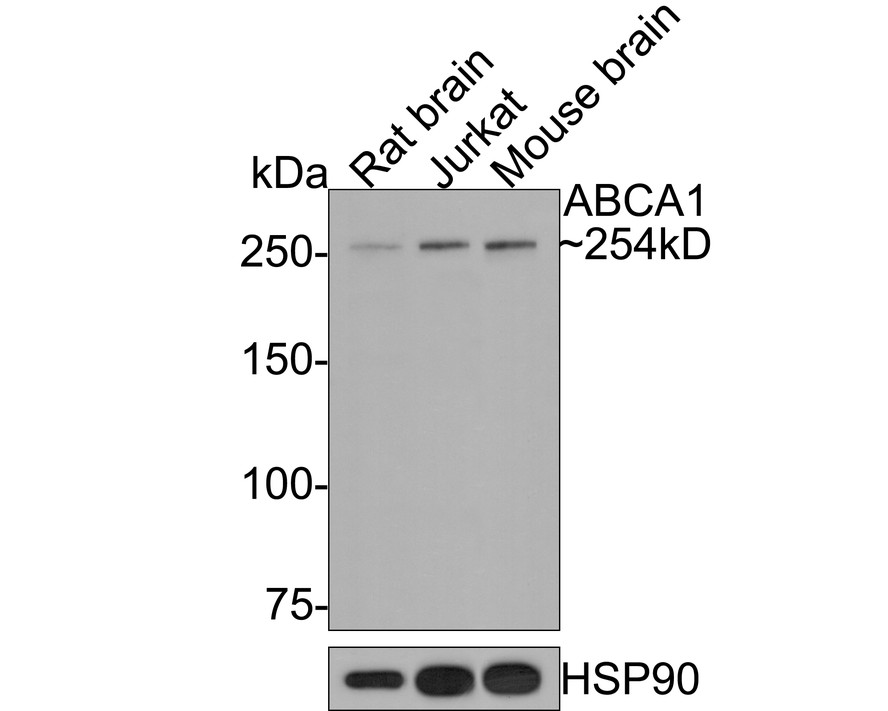
Fig1: Western blot analysis of ABCA1 on different lysates with Rabbit anti-ABCA1 antibody at 1/500 dilution. Lane 1: Rat brain tissue lysate (20 µg/Lane), Lane 2: Jurkat cell lysate (10 µg/Lane), Lane 3: Mouse brain tissue lysate (20 µg/Lane), Exposure time: 2 minutes; 6% SDS-PAGE gel. Proteins were transferred to a PVDF membrane and blocked with 5% NFDM/TBST for 1 hour at room temperature. The primary antibody at 1/500 dilution was used in 5% NFDM/TBST at room temperature for 2 hours. Goat Anti-Rabbit IgG - HRP Secondary Antibody at 1:300,000 dilution was used for 1 hour at room temperature.Application
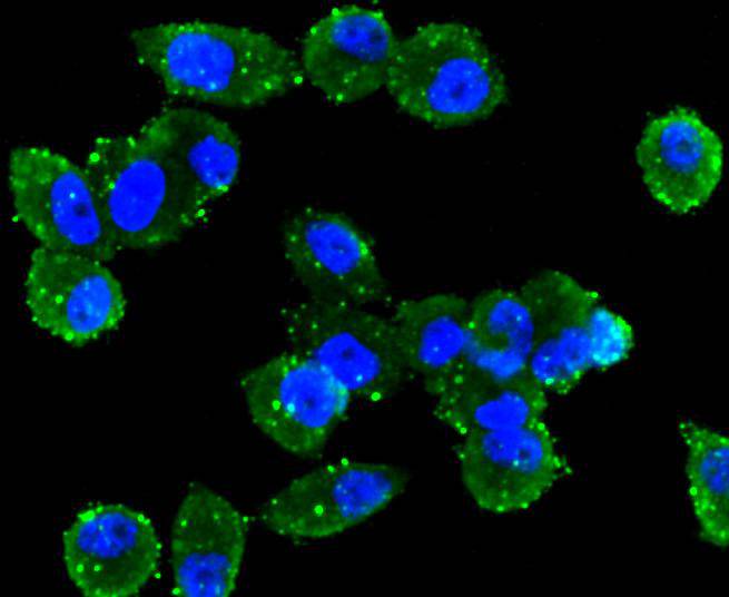
Fig2: Immunocytochemical staining of PC-3M cells using anti-ABCA1 rabbit polyclonal antibody.Application
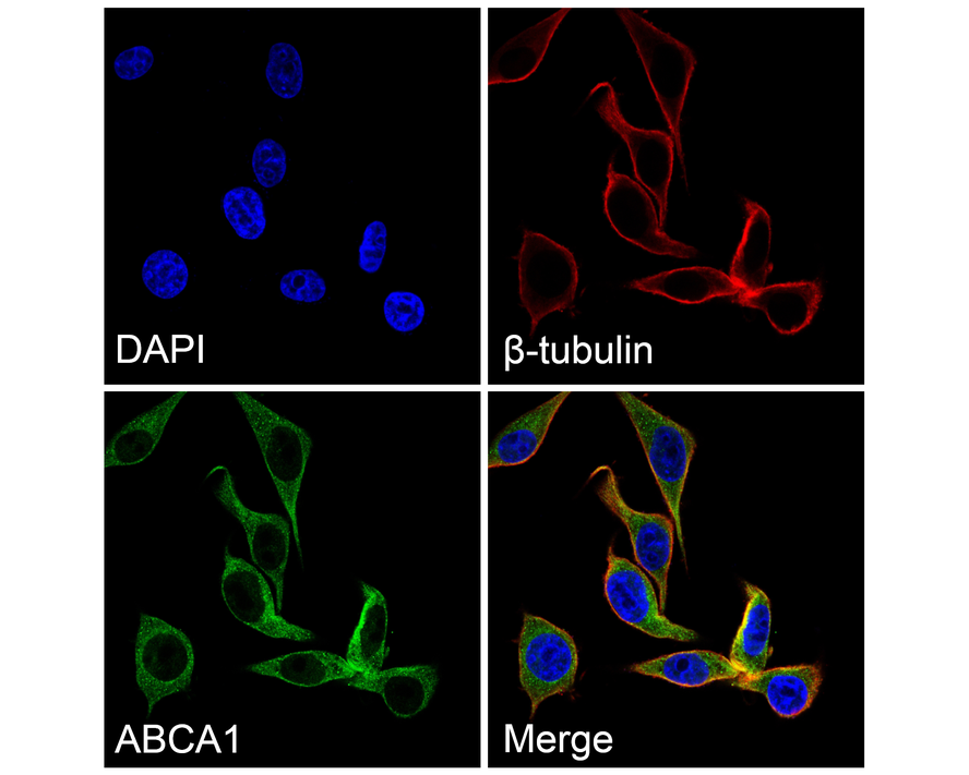
Fig3: Immunocytochemistry analysis of HepG2 cells labeling ABCA1 with Rabbit anti-ABCA1 antibody at 1/200 dilution. Cells were fixed in 4% paraformaldehyde for 10 minutes at 37 ℃, permeabilized with 0.05% Triton X-100 in PBS for 20 minutes, and then blocked with 2% negative goat serum for 30 minutes at room temperature. Cells were then incubated with Rabbit anti-ABCA1 antibody at 1/200 dilution in 2% negative goat serum overnight at 4 ℃. Goat Anti-Rabbit IgG H&L (488) was used as the secondary antibody at 1/1,000 dilution. Nuclear DNA was labelled in blue with DAPI. β-tubulin (red) was stained at 1/100 dilution overnight at +4℃. Goat Anti-Mouse IgG H&L (647) were used as the secondary antibody at 1/1,000 dilution.Application
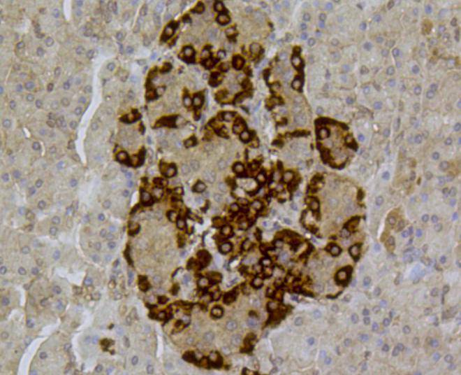
Fig4: Immunohistochemical analysis of paraffin- embedded human pancreas tissue using anti-ABCA1 rabbit polyclonal antibody.Application
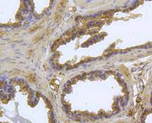
Fig5: Immunohistochemical analysis of paraffin- embedded mouse prostate tissue using anti-ABCA1 rabbit polyclonal antibody.Application
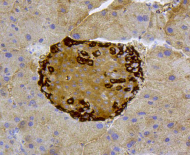
Fig6: Immunohistochemical analysis of paraffin- embedded mouse pancreas tissue using anti-ABCA1 rabbit polyclonal antibody.Application
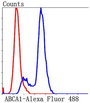
Fig7: Flow cytometric analysis of Jurkat cells with ABCA1 antibody at 1/50 dilution (blue) compared with an unlabelled control (cells without incubation with primary antibody; red). Alexa Fluor 488-conjugated Goat anti rabbit IgG was used as the secondary antibody.| Product Name | ABCA1 Rabbit Polyclonal Antibody |
|---|---|
| Antibody Type | Primary Antibodies |
| Product description | ABC1 (for ATP-binding cassette transporter 1) is a member of the family of ATP-binding cassette proteins which transport various molecules across biological membranes. ABC1 contains two characteristic ATP-binding domains and 12 transmembrane domains which form a channel-like structure for transport. Mutations in the ABC1 gene are implicated in Tangier disease, characterized by low serum high density lipoprotein. ABC1 is widely expressed in human tissues, with high levels of expression in liver, lung, adrenal glands, placenta and fetal tissue. ABC1 expression is induced during monocyte differentiation and upregulated in the presence of acetylated low-density lipoprotein. ABC1 may have a dual regulatory function in macrophage lipid metabolism and inflammation. |
| Immunogen | Synthetic peptide within Human ABCA1 aa 21-70 / 2,261. |
| Clonality | Polyclonal |
|---|---|
| Isotype | IgG |
| Host Species | Rabbit |
| Tested Applications | FCICC/IFIHCWB |
| WB:1:500-1:1000 ICC/IF:1:50-1:200 IHC:1:50-1:200 FC:1:50-1:100 |
|
| Species Reactivity | HumanMouseRat |
| Concentration | 1mg/ml |
| Purification | Affinity purified |
| Alternative Names | ABC 1 antibody ABC Transporter 1 antibody ABC-1 antibody ABC1 antibody ABCA 1 antibody ABCA1 antibody ABCA1_HUMAN antibody ATP binding Cassette 1 antibody ATP binding cassette sub family A ABC1 member 1 antibody ATP binding cassette sub family A member 1 antibody ATP binding Cassette Transporter 1 antibody ATP-binding cassette 1 antibody ATP-binding cassette sub-family A member 1 antibody ATP-binding cassette transporter 1 antibody CERP antibody Cholesterol efflux regulatory protein antibody FLJ14958 antibody HDLDT1 antibody Membrane bound antibody MGC164864 antibody MGC165011 antibody TD antibody TGD antibody |
|---|---|
| Molecular Weight(MW) | 254 kDa |
| Cellular Localization | Membrane |
| SwissProt ID | O95477 |
|---|
Application
Fig1: Western blot analysis of ABCA1 on different lysates with Rabbit anti-ABCA1 antibody at 1/500 dilution. Lane 1: Rat brain tissue lysate (20 µg/Lane), Lane 2: Jurkat cell lysate (10 µg/Lane), Lane 3: Mouse brain tissue lysate (20 µg/Lane), Exposure time: 2 minutes; 6% SDS-PAGE gel. Proteins were transferred to a PVDF membrane and blocked with 5% NFDM/TBST for 1 hour at room temperature. The primary antibody at 1/500 dilution was used in 5% NFDM/TBST at room temperature for 2 hours. Goat Anti-Rabbit IgG - HRP Secondary Antibody at 1:300,000 dilution was used for 1 hour at room temperature.
Application
Fig2: Immunocytochemical staining of PC-3M cells using anti-ABCA1 rabbit polyclonal antibody.
Application
Fig3: Immunocytochemistry analysis of HepG2 cells labeling ABCA1 with Rabbit anti-ABCA1 antibody at 1/200 dilution. Cells were fixed in 4% paraformaldehyde for 10 minutes at 37 ℃, permeabilized with 0.05% Triton X-100 in PBS for 20 minutes, and then blocked with 2% negative goat serum for 30 minutes at room temperature. Cells were then incubated with Rabbit anti-ABCA1 antibody at 1/200 dilution in 2% negative goat serum overnight at 4 ℃. Goat Anti-Rabbit IgG H&L (488) was used as the secondary antibody at 1/1,000 dilution. Nuclear DNA was labelled in blue with DAPI. β-tubulin (red) was stained at 1/100 dilution overnight at +4℃. Goat Anti-Mouse IgG H&L (647) were used as the secondary antibody at 1/1,000 dilution.
Application
Fig4: Immunohistochemical analysis of paraffin- embedded human pancreas tissue using anti-ABCA1 rabbit polyclonal antibody.
Application
Fig5: Immunohistochemical analysis of paraffin- embedded mouse prostate tissue using anti-ABCA1 rabbit polyclonal antibody.
Application
Fig6: Immunohistochemical analysis of paraffin- embedded mouse pancreas tissue using anti-ABCA1 rabbit polyclonal antibody.
Application
Fig7: Flow cytometric analysis of Jurkat cells with ABCA1 antibody at 1/50 dilution (blue) compared with an unlabelled control (cells without incubation with primary antibody; red). Alexa Fluor 488-conjugated Goat anti rabbit IgG was used as the secondary antibody.| Application Notes | WB:1:500-1:1000 ICC/IF:1:50-1:200 IHC:1:50-1:200 FC:1:50-1:100 |
|---|
| Form | Liquid |
|---|---|
| Storage Instructions | Store at +4℃ after thawing. Aliquot store at -20℃ or -80℃. Avoid repeated freeze / thaw cycles. |
| Storage Buffer | 1*TBS (pH7.4), 1%BSA, 40%Glycerol. Preservative: 0.05% Sodium Azide. |


