Application
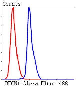
Fig11: Flow cytometric analysis of HepG2 cells with BECN1 antibody at 1/50 dilution (blue) compared with an unlabelled control (cells without incubation with primary antibody; red). Alexa Fluor 488-conjugated Goat anti rabbit IgG was used as the secondary antibody.Application
Fig11: Flow cytometric analysis of HepG2 cells with BECN1 antibody at 1/50 dilution (blue) compared with an unlabelled control (cells without incubation with primary antibody; red). Alexa Fluor 488-conjugated Goat anti rabbit IgG was used as the secondary antibody.Application
Fig11: Flow cytometric analysis of HepG2 cells with BECN1 antibody at 1/50 dilution (blue) compared with an unlabelled control (cells without incubation with primary antibody; red). Alexa Fluor 488-conjugated Goat anti rabbit IgG was used as the secondary antibody.Application
Fig11: Flow cytometric analysis of HepG2 cells with BECN1 antibody at 1/50 dilution (blue) compared with an unlabelled control (cells without incubation with primary antibody; red). Alexa Fluor 488-conjugated Goat anti rabbit IgG was used as the secondary antibody.Application
Fig11: Flow cytometric analysis of HepG2 cells with BECN1 antibody at 1/50 dilution (blue) compared with an unlabelled control (cells without incubation with primary antibody; red). Alexa Fluor 488-conjugated Goat anti rabbit IgG was used as the secondary antibody.Application
Fig11: Flow cytometric analysis of HepG2 cells with BECN1 antibody at 1/50 dilution (blue) compared with an unlabelled control (cells without incubation with primary antibody; red). Alexa Fluor 488-conjugated Goat anti rabbit IgG was used as the secondary antibody.Application
Fig11: Flow cytometric analysis of HepG2 cells with BECN1 antibody at 1/50 dilution (blue) compared with an unlabelled control (cells without incubation with primary antibody; red). Alexa Fluor 488-conjugated Goat anti rabbit IgG was used as the secondary antibody.Application
Fig11: Flow cytometric analysis of HepG2 cells with BECN1 antibody at 1/50 dilution (blue) compared with an unlabelled control (cells without incubation with primary antibody; red). Alexa Fluor 488-conjugated Goat anti rabbit IgG was used as the secondary antibody.Application
Fig11: Flow cytometric analysis of HepG2 cells with BECN1 antibody at 1/50 dilution (blue) compared with an unlabelled control (cells without incubation with primary antibody; red). Alexa Fluor 488-conjugated Goat anti rabbit IgG was used as the secondary antibody.Application
Fig11: Flow cytometric analysis of HepG2 cells with BECN1 antibody at 1/50 dilution (blue) compared with an unlabelled control (cells without incubation with primary antibody; red). Alexa Fluor 488-conjugated Goat anti rabbit IgG was used as the secondary antibody.Application
Fig11: Flow cytometric analysis of HepG2 cells with BECN1 antibody at 1/50 dilution (blue) compared with an unlabelled control (cells without incubation with primary antibody; red). Alexa Fluor 488-conjugated Goat anti rabbit IgG was used as the secondary antibody.| Product Name | BECN1 |
|---|---|
| Antibody Type | Primary Antibodies |
| Antigen Alias | APG6 antibody ATG 6 antibody ATG6 antibody ATG6 autophagy related 6 homolog antibody Bcl-2-interacting protein beclin antibody Beclin 1 (coiled coil moesin like BCL2 interacting protein) antibody Beclin 1 autophagy related antibody Beclin-1 antibody Beclin1 antibody BECN 1 antibody Becn1 antibody BECN1_HUMAN antibody Coiled coil myosin like BCL2 interacting protein antibody Coiled-coil myosin-like BCL2-interacting protein antibody GT197 antibody Protein GT197 antibody VPS 30 antibody VPS30 antibody |
| Product description | BECN1 (Beclin 1) is a coiled-coil protein that has been implicated as an inhibitor of tumorigenesis. BECN1, which associates with Bcl-2, plays a significant role in autophagy. Autophagy is the degradation of cellular proteins in the lysosomes, and when this pathway is suppressed, cell growth is deregulated. Autophagy is controlled by the same signal transduction pathway that induces the phosphorylation of the Ribosomal Protein S6, and both are mediated via amino acids. BECN1 expression in various carcinoma cell lines such as MCF7 is low, whereas it is ubiquitously expressed in normal breast tissue. In transfected MCF7 cells, BECN1 complements autophagocytosis and, subsequently, inhibits cellular proliferation. Additionally, BECN1 shares structural similarity to the yeast autophagy gene product, Apg6, and was one of the first mammalian proteins discovered to mediate autophagy. |
| Immunogen | Synthetic peptide (KLH-coupled) within human BECN1 C terminal. |
| Clonality | Polyclonal |
|---|---|
| Isotype | IgG |
| Host Species | Rabbit |
| Tested Applications | ICCIHCFC |
| ICC:1:50-1:200 IHC:1:50-1:200 FC:1:50-1:100 |
|
| Species Reactivity | HumanMouse |
| Concentration | 1mg/ml |
| Alternative Names | APG6 antibody ATG 6 antibody ATG6 antibody ATG6 autophagy related 6 homolog antibody Bcl-2-interacting protein beclin antibody Beclin 1 (coiled coil moesin like BCL2 interacting protein) antibody Beclin 1 autophagy related antibody Beclin-1 antibody Beclin1 antibody BECN 1 antibody Becn1 antibody BECN1_HUMAN antibody Coiled coil myosin like BCL2 interacting protein antibody Coiled-coil myosin-like BCL2-interacting protein antibody GT197 antibody Protein GT197 antibody VPS 30 antibody VPS30 antibody |
|---|---|
| Molecular Weight(MW) | 52 kDa |
| Cellular Localization | Cytoplasm, Endosome, Mitochondrion membrane Mitochondrion membrane, Endoplasmic reticulum membrane |
Application
Fig11: Flow cytometric analysis of HepG2 cells with BECN1 antibody at 1/50 dilution (blue) compared with an unlabelled control (cells without incubation with primary antibody; red). Alexa Fluor 488-conjugated Goat anti rabbit IgG was used as the secondary antibody.
Application
Fig11: Flow cytometric analysis of HepG2 cells with BECN1 antibody at 1/50 dilution (blue) compared with an unlabelled control (cells without incubation with primary antibody; red). Alexa Fluor 488-conjugated Goat anti rabbit IgG was used as the secondary antibody.
Application
Fig11: Flow cytometric analysis of HepG2 cells with BECN1 antibody at 1/50 dilution (blue) compared with an unlabelled control (cells without incubation with primary antibody; red). Alexa Fluor 488-conjugated Goat anti rabbit IgG was used as the secondary antibody.
Application
Fig11: Flow cytometric analysis of HepG2 cells with BECN1 antibody at 1/50 dilution (blue) compared with an unlabelled control (cells without incubation with primary antibody; red). Alexa Fluor 488-conjugated Goat anti rabbit IgG was used as the secondary antibody.
Application
Fig11: Flow cytometric analysis of HepG2 cells with BECN1 antibody at 1/50 dilution (blue) compared with an unlabelled control (cells without incubation with primary antibody; red). Alexa Fluor 488-conjugated Goat anti rabbit IgG was used as the secondary antibody.
Application
Fig11: Flow cytometric analysis of HepG2 cells with BECN1 antibody at 1/50 dilution (blue) compared with an unlabelled control (cells without incubation with primary antibody; red). Alexa Fluor 488-conjugated Goat anti rabbit IgG was used as the secondary antibody.
Application
Fig11: Flow cytometric analysis of HepG2 cells with BECN1 antibody at 1/50 dilution (blue) compared with an unlabelled control (cells without incubation with primary antibody; red). Alexa Fluor 488-conjugated Goat anti rabbit IgG was used as the secondary antibody.
Application
Fig11: Flow cytometric analysis of HepG2 cells with BECN1 antibody at 1/50 dilution (blue) compared with an unlabelled control (cells without incubation with primary antibody; red). Alexa Fluor 488-conjugated Goat anti rabbit IgG was used as the secondary antibody.
Application
Fig11: Flow cytometric analysis of HepG2 cells with BECN1 antibody at 1/50 dilution (blue) compared with an unlabelled control (cells without incubation with primary antibody; red). Alexa Fluor 488-conjugated Goat anti rabbit IgG was used as the secondary antibody.
Application
Fig11: Flow cytometric analysis of HepG2 cells with BECN1 antibody at 1/50 dilution (blue) compared with an unlabelled control (cells without incubation with primary antibody; red). Alexa Fluor 488-conjugated Goat anti rabbit IgG was used as the secondary antibody.
Application
Fig11: Flow cytometric analysis of HepG2 cells with BECN1 antibody at 1/50 dilution (blue) compared with an unlabelled control (cells without incubation with primary antibody; red). Alexa Fluor 488-conjugated Goat anti rabbit IgG was used as the secondary antibody.| Positive Control | Hela, N2A, MCF-7, SHG-44, human liver cancer tissue, human breast tissue, human lung tissue, human kidney tissue, mouse brain tissue, mouse heart tissue, HepG2. |
|---|---|
| Application Notes | ICC:1:50-1:200 IHC:1:50-1:200 FC:1:50-1:100 |
| Form | Liquid |
|---|---|
| Storage Instructions | Store at +4℃ after thawing. Aliquot store at -20℃. Avoid repeated freeze / thaw cycles. |
| Storage Buffer | 1*TBS (pH7.4), 1%BSA, 40%Glycerol. Preservative: 0.05% Sodium Azide. |


