Application
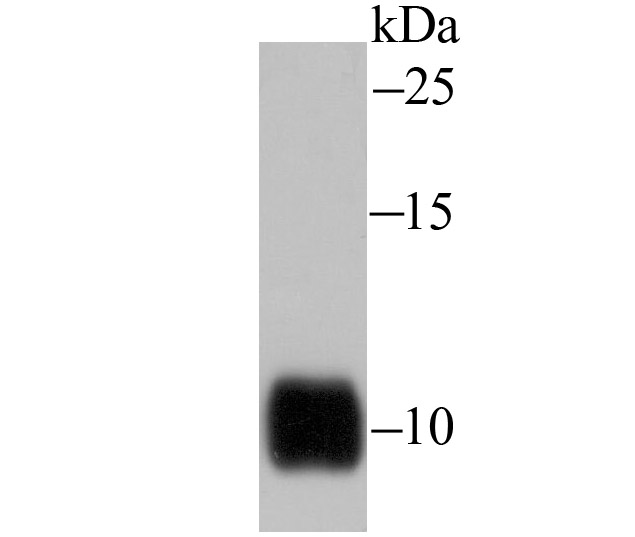
Fig1: Western blot analysis of IL-8 on human kidney tissue lysate using anti-IL-8 antibody at 1/1,000 dilution.Application
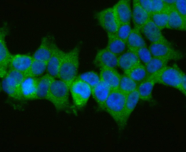
Fig2: ICC staining IL-8 in 293T cells (green). The nuclear counter stain is DAPI (blue). Cells were fixed in paraformaldehyde, permeabilised with 0.25% Triton X100/PBS.Application
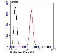
Fig3: Flow cytometric analysis of HUVEC cells with IL-8 antibody at 1/100 dilution (red) compared with an unlabelled control (cells without incubation with primary antibody; black). Alexa Fluor 488-conjugated goat anti-rabbit IgG was used as the secondary antibody.| Product Name | IL-8 |
|---|---|
| Antibody Type | Primary Antibodies |
| Antigen Alias | (Ala-IL-8)77 antibody (Ser-IL-8)72 antibody 9E3 antibody Beta thromboglobulin like protein antibody C-X-C motif chemokine 8 antibody CEF-4 antibody chemokine, CXC motif, ligand 8 antibody CXCL8 antibody Emoctakin antibody GCP-1 antibody GCP/IL-8 protein I antibody GCP/IL-8 protein II antibody GCP/IL-8 protein III antibody GCP/IL-8 protein IV antibody GCP/IL-8 protein V antibody GCP/IL-8 protein VI antibody GCP1 antibody Granulocyte chemotactic protein 1 antibody IL-8 antibody IL-8(1-77) antibody IL-8(9-77) antibody IL8 antibody IL8/NAP1 form I antibody IL8/NAP1 form II antibody IL8/NAP1 form III antibody IL8/NAP1 form IV antibody IL8/NAP1 form V antibody IL8/NAP1 form VI antibody IL8_HUMAN antibody Inteleukin 8 antibody LECT antibody LUCT antibody Lymphocyte-derived neutrophil-activating factor antibody LYNAP antibody MDNCF antibody MDNCF-b antibody MDNCF-c antibody MONAP antibody Monocyte-derived neutrophil chemotactic factor antibody Monocyte-derived neutrophil-activating peptide antibody NAF antibody NAP-1 antibody NAP1 antibody Neutrophil activating peptide 1 antibody Neutrophil activating protein 1 antibody Neutrophil-activating factor antibody Neutrophil-activating protein 1 antibody Protein 3 10C antibody Protein 3-10C antibody SCYB8 antibody Small inducible cytokine subfamily B member 8 antibody T cell chemotactic factor antibody T-cell chemotactic factor antibody TSG1 antibody |
| Product description | Interleukin-8, or IL-8, the prototypic member of the C-X-C, or a, family of chemokines, is a chemoattractant cytokine involved in the chemotaxis and activation of neutrophils. IL-8 expression has been correlated to a large number of chronic inflammatory diseases, including inflammatory bowel disease (IBD) and atherosclerosis. IL-8 is cleaved from a 99 amino acid precursor to a 72 amino acid, nonglycosylated, biologically active protein. IL-8 monomers and dimers exhibit a dynamic equilibrium both free in solution and in cell surface-bound forms and thus regulate chemotaxis and receptor signaling. Research has shown that IL-8 dimerization functions as a negative regulator for IL-8 receptor function. Two IL-8 receptors, designated IL-8RA and IL-8RB, have been described and share 77% sequence identity. Both are seven-transmembrane domain proteins (7TMD), similar to the G protein-coupled receptors and, in addition to IL-8, serve as receptors for other members of the a and b chemokine families. |
| Immunogen | Recombinant full length protein of human IL-8. |
| Clonality | Polyclonal |
|---|---|
| Isotype | IgG |
| Host Species | Rabbit |
| Tested Applications | WBICCFC |
| WB:1:500-1:2,000 ICC:1:500-1:1,000 FC:1:50-1:100 |
|
| Species Reactivity | Human |
| Concentration | 1mg/ml |
| Alternative Names | (Ala-IL-8)77 antibody
(Ser-IL-8)72 antibody
9E3 antibody
Beta thromboglobulin like protein antibody
C-X-C motif chemokine 8 antibody
CEF-4 antibody
chemokine CXC motif ligand 8 antibody CXCL8 antibody Emoctakin antibody GCP-1 antibody GCP/IL-8 protein I antibody GCP/IL-8 protein II antibody GCP/IL-8 protein III antibody GCP/IL-8 protein IV antibody GCP/IL-8 protein V antibody GCP/IL-8 protein VI antibody GCP1 antibody Granulocyte chemotactic protein 1 antibody IL-8 antibody IL-8(1-77) antibody IL-8(9-77) antibody IL8 antibody IL8/NAP1 form I antibody IL8/NAP1 form II antibody IL8/NAP1 form III antibody IL8/NAP1 form IV antibody IL8/NAP1 form V antibody IL8/NAP1 form VI antibody IL8_HUMAN antibody Inteleukin 8 antibody LECT antibody LUCT antibody Lymphocyte-derived neutrophil-activating factor antibody LYNAP antibody MDNCF antibody MDNCF-b antibody MDNCF-c antibody MONAP antibody Monocyte-derived neutrophil chemotactic factor antibody Monocyte-derived neutrophil-activating peptide antibody NAF antibody NAP-1 antibody NAP1 antibody Neutrophil activating peptide 1 antibody Neutrophil activating protein 1 antibody Neutrophil-activating factor antibody Neutrophil-activating protein 1 antibody Protein 3 10C antibody Protein 3-10C antibody SCYB8 antibody Small inducible cytokine subfamily B member 8 antibody T cell chemotactic factor antibody T-cell chemotactic factor antibody TSG1 antibody |
|---|---|
| Molecular Weight(MW) | 11 kDa |
| Cellular Localization | Secreted. |
| SwissProt ID | P10145 |
|---|
Application
Fig1: Western blot analysis of IL-8 on human kidney tissue lysate using anti-IL-8 antibody at 1/1,000 dilution.
Application
Fig2: ICC staining IL-8 in 293T cells (green). The nuclear counter stain is DAPI (blue). Cells were fixed in paraformaldehyde, permeabilised with 0.25% Triton X100/PBS.
Application
Fig3: Flow cytometric analysis of HUVEC cells with IL-8 antibody at 1/100 dilution (red) compared with an unlabelled control (cells without incubation with primary antibody; black). Alexa Fluor 488-conjugated goat anti-rabbit IgG was used as the secondary antibody.| Positive Control | Human kidney tissue, 293T, HUVEC. |
|---|---|
| Application Notes | WB:1:500-1:2,000 ICC:1:500-1:1,000 FC:1:50-1:100 |
| Form | Liquid |
|---|---|
| Storage Instructions | Store at +4℃ after thawing. Aliquot store at -20℃. Avoid repeated freeze / thaw cycles. |
| Storage Buffer | 1*TBS (pH7.4), 0.5%BSA, 50%Glycerol. Preservative: 0.05% Sodium Azide. |


