Application
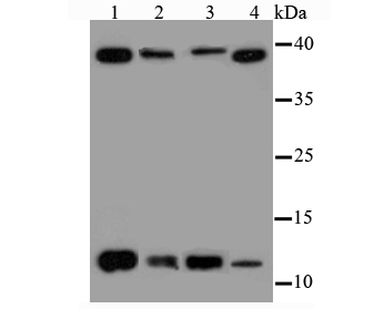
Fig1: Western blot analysis of Thioredoxin on different cell lysates using anti-Thioredoxin antibody at 1/500 dilution. Positive control: Lane1: Hela Lane2: HepG2 Lane3: A431 Lane4: PC-3MApplication
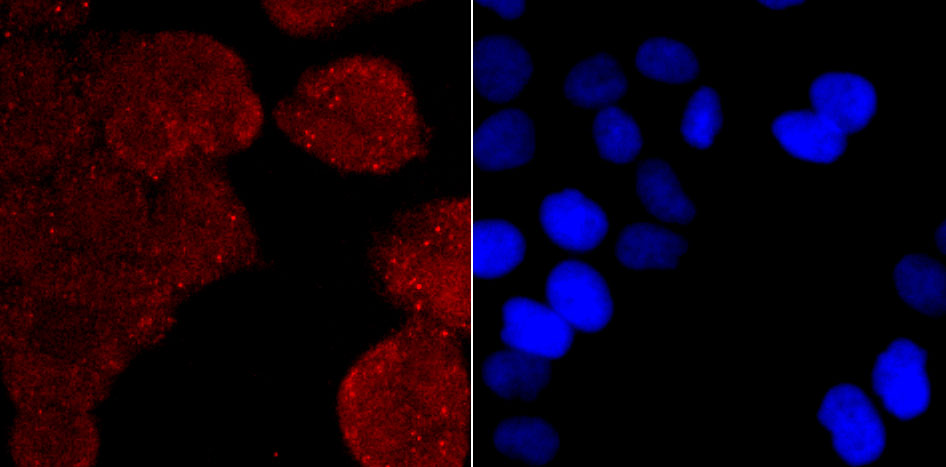
Fig2: ICC staining Thioredoxin in Hela cells (red). The nuclear counter stain is DAPI (blue). Cells were fixed in paraformaldehyde, permeabilised with 0.25% Triton X100/PBS.Application
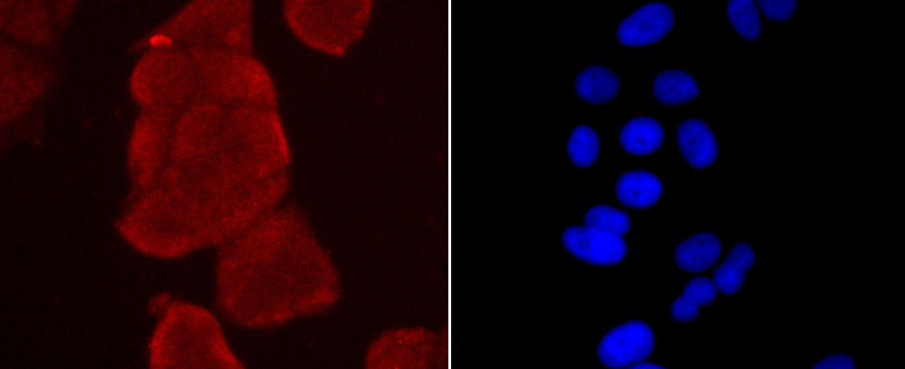
Fig3: ICC staining Thioredoxin in MCF-7 cells (red). The nuclear counter stain is DAPI (blue). Cells were fixed in paraformaldehyde, permeabilised with 0.25% Triton X100/PBS.Application
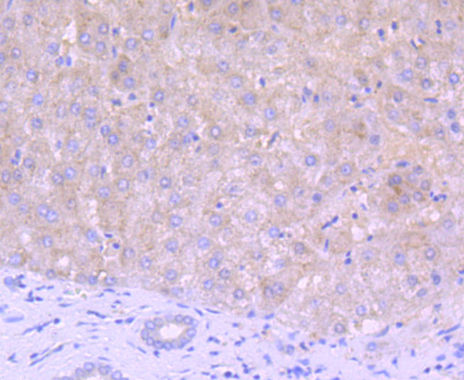
Fig4: Immunohistochemical analysis of paraffin-embedded human liver tissue using anti-Thioredoxin antibody. Counter stained with hematoxylin.Application
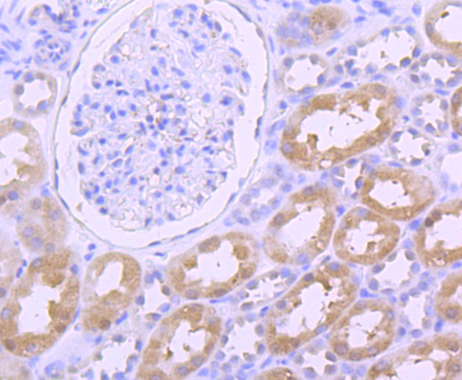
Fig5: Immunohistochemical analysis of paraffin-embedded human kidney tissue using anti-Thioredoxin antibody. Counter stained with hematoxylin.Application
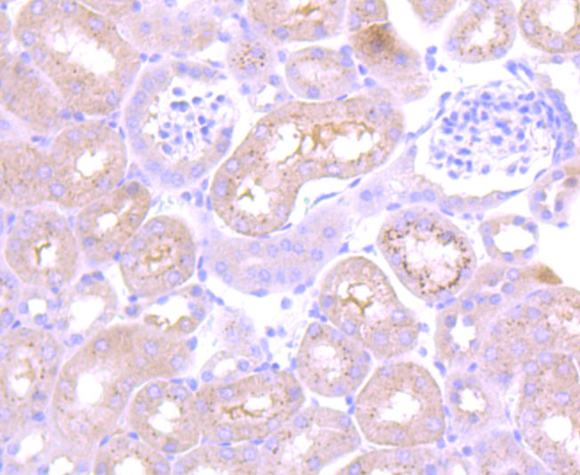
Fig6: Immunohistochemical analysis of paraffin-embedded mouse kidney tissue using anti-Thioredoxin antibody. Counter stained with hematoxylin.Application
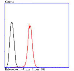
Fig7: Flow cytometric analysis of Hela cells with Thioredoxin antibody at 1/100 dilution (red) compared with an unlabelled control (cells without incubation with primary antibody; black).| Product Name | Thioredoxin |
|---|---|
| Antibody Type | Primary Antibodies |
| Antigen Alias | ADF antibody ATL-derived factor antibody SASP antibody Surface associated sulphydryl protein antibody TRN antibody TXN 1 antibody TXN antibody |
| Product description | Participates in various redox reactions through the reversible oxidation of its active center dithiol to a disulfide and catalyzes dithiol-disulfide exchange reactions. Plays a role in the reversible S-nitrosylation of cysteine residues in target proteins, and thereby contributes to the response to intracellular nitric oxide. Nitrosylates the active site Cys of CASP3 in response to nitric oxide (NO), and thereby inhibits caspase-3 activity. Induces the FOS/JUN AP-1 DNA-binding activity in ionizing radiation (IR) cells through its oxidation/reduction status and stimulates AP-1 transcriptional activity.ADF augments the expression of the interleukin-2 receptor TAC (IL2R/P55). |
| Immunogen | Peptide |
| Clonality | Polyclonal |
|---|---|
| Isotype | IgG |
| Host Species | Rabbit |
| Tested Applications | WBICCIHC |
| WB:1:500 ICC:1:50-1:200 IHC:1:50-1:100 FC:1:50-1:100 |
|
| Species Reactivity | HumanMouse |
| Concentration | 1mg/ml |
| Alternative Names | ADF antibody ATL-derived factor antibody SASP antibody Surface associated sulphydryl protein antibody TRN antibody TXN 1 antibody TXN antibody |
|---|---|
| Molecular Weight(MW) | 12 kDa, additional band 40 kDa. |
| Cellular Localization | Nucleus. Cytoplasm. Secreted. |
Application
Fig1: Western blot analysis of Thioredoxin on different cell lysates using anti-Thioredoxin antibody at 1/500 dilution. Positive control: Lane1: Hela Lane2: HepG2 Lane3: A431 Lane4: PC-3M
Application
Fig2: ICC staining Thioredoxin in Hela cells (red). The nuclear counter stain is DAPI (blue). Cells were fixed in paraformaldehyde, permeabilised with 0.25% Triton X100/PBS.
Application
Fig3: ICC staining Thioredoxin in MCF-7 cells (red). The nuclear counter stain is DAPI (blue). Cells were fixed in paraformaldehyde, permeabilised with 0.25% Triton X100/PBS.
Application
Fig4: Immunohistochemical analysis of paraffin-embedded human liver tissue using anti-Thioredoxin antibody. Counter stained with hematoxylin.
Application
Fig5: Immunohistochemical analysis of paraffin-embedded human kidney tissue using anti-Thioredoxin antibody. Counter stained with hematoxylin.
Application
Fig6: Immunohistochemical analysis of paraffin-embedded mouse kidney tissue using anti-Thioredoxin antibody. Counter stained with hematoxylin.
Application
Fig7: Flow cytometric analysis of Hela cells with Thioredoxin antibody at 1/100 dilution (red) compared with an unlabelled control (cells without incubation with primary antibody; black).| Positive Control | Hela, HepG2, A431, PC-3M, MCF-7, human liver tissue, human kidney tissue, mouse kidney tissue. |
|---|---|
| Application Notes | WB:1:500 ICC:1:50-1:200 IHC:1:50-1:100 FC:1:50-1:100 |
| Form | Liquid |
|---|---|
| Storage Instructions | Store at +4℃ after thawing. Aliquot store at -20℃ or -80℃. Avoid repeated freeze / thaw cycles. |
| Storage Buffer | 1*TBS (pH7.4), 1%BSA, 40%Glycerol. Preservative: 0.05% Sodium Azide. |


