Application
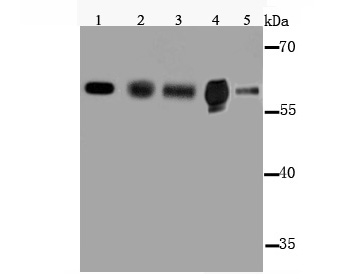
Fig1: Western blot analysis of Cytochrome P450 2E1 on different lysates using anti-Cytochrome P450 2E1 antibody at 1/1000 dilution. Positive control: Lane 1: MCF-7 Lane 2: NIH-3T3 Lane 3: Mouse kidney tissue Lane 4: siHaApplication
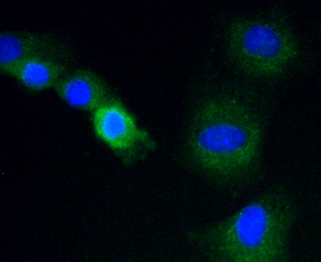
Fig2: ICC staining Cytochrome P450 2E1 in C2C12 cells (green). The nuclear counter stain is DAPI (blue). Cells were fixed in paraformaldehyde, permeabilised with 0.25% Triton X100/PBS.Application
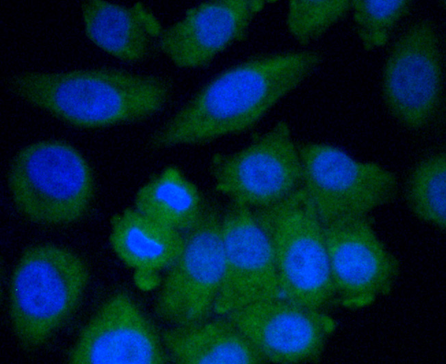
Fig3: ICC staining Cytochrome P450 2E1 in HepG2 cells (green). The nuclear counter stain is DAPI (blue). Cells were fixed in paraformaldehyde, permeabilised with 0.25% Triton X100/PBS.Application
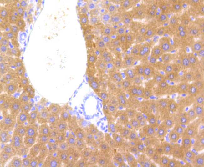
Fig4: Immunohistochemical analysis of paraffin-embedded mouse liver tissue using anti-Cytochrome P450 2E1 antibody. Counter stained with hematoxylin.| Product Name | Cytochrome P450 2E1 |
|---|---|
| Antibody Type | Primary Antibodies |
| Antigen Alias | 4-nitrophenol 2-hydroxylase antibody CP2E1_HUMAN antibody CPE1 antibody CYP2E antibody Cyp2e1 antibody CYPIIE1 antibody Cytochrome P450 2E1 antibody Cytochrome P450 isozyme 3A antibody Cytochrome P450 LM3A antibody Cytochrome P450, family 2, subfamily e, polypeptide 1 antibody cytochrome P450, subfamily IIE (ethanol-inducible), polypeptide 1 antibody Cytochrome P450, subfamily IIE antibody Cytochrome P450-ALC antibody Cytochrome P450-J antibody Cytochrome P450RLM6 antibody EC 1.14.13.- antibody EC 1.14.13.n7 antibody Ethanol-inducible P450 antibody Flavoprotein-linked monooxygenase antibody MGC133529 antibody Microsomal monooxygenase antibody OTTHUMP00000020813 antibody OTTHUMP00000046571 antibody P450 ALC antibody P450-J antibody P450C2E antibody P450J antibody P450RLM6 antibody Xenobiotic monooxygenase antibody |
| Product description | Metabolizes several precarcinogens, drugs, and solvents to reactive metabolites. Inactivates a number of drugs and xenobiotics and also bioactivates many xenobiotic substrates to their hepatotoxic or carcinogenic forms. |
| Immunogen | Peptide |
| Clonality | Polyclonal |
|---|---|
| Isotype | IgG |
| Host Species | Rabbit |
| Tested Applications | WBICCIHC |
| WB:1:1000 ICC:1:50-1:200 IHC:1:50-1:200 |
|
| Species Reactivity | HumanMouse |
| Concentration | 1mg/ml |
| Alternative Names | 4-nitrophenol 2-hydroxylase antibody
CP2E1_HUMAN antibody
CPE1 antibody
CYP2E antibody
Cyp2e1 antibody
CYPIIE1 antibody
Cytochrome P450 2E1 antibody
Cytochrome P450 isozyme 3A antibody
Cytochrome P450 LM3A antibody
Cytochrome P450 family 2 subfamily e polypeptide 1 antibody cytochrome P450 subfamily IIE (ethanol-inducible) polypeptide 1 antibody Cytochrome P450 subfamily IIE antibody Cytochrome P450-ALC antibody Cytochrome P450-J antibody Cytochrome P450RLM6 antibody EC 1.14.13.- antibody EC 1.14.13.n7 antibody Ethanol-inducible P450 antibody Flavoprotein-linked monooxygenase antibody MGC133529 antibody Microsomal monooxygenase antibody OTTHUMP00000020813 antibody OTTHUMP00000046571 antibody P450 ALC antibody P450-J antibody P450C2E antibody P450J antibody P450RLM6 antibody Xenobiotic monooxygenase antibody |
|---|---|
| Molecular Weight(MW) | 57 kDa |
| Cellular Localization | Endoplasmic reticulum membrane. Microsome membrane. |
Application
Fig1: Western blot analysis of Cytochrome P450 2E1 on different lysates using anti-Cytochrome P450 2E1 antibody at 1/1000 dilution. Positive control: Lane 1: MCF-7 Lane 2: NIH-3T3 Lane 3: Mouse kidney tissue Lane 4: siHa
Application
Fig2: ICC staining Cytochrome P450 2E1 in C2C12 cells (green). The nuclear counter stain is DAPI (blue). Cells were fixed in paraformaldehyde, permeabilised with 0.25% Triton X100/PBS.
Application
Fig3: ICC staining Cytochrome P450 2E1 in HepG2 cells (green). The nuclear counter stain is DAPI (blue). Cells were fixed in paraformaldehyde, permeabilised with 0.25% Triton X100/PBS.
Application
Fig4: Immunohistochemical analysis of paraffin-embedded mouse liver tissue using anti-Cytochrome P450 2E1 antibody. Counter stained with hematoxylin.| Positive Control | MCF-7, NIH-3T3, mouse kidney tissue lysate, C2C12, HepG2, mouse liver tissue. |
|---|---|
| Application Notes | WB:1:1000 ICC:1:50-1:200 IHC:1:50-1:200 |
| Form | Liquid |
|---|---|
| Storage Instructions | Store at +4℃ after thawing. Aliquot store at -20℃ or -80℃. Avoid repeated freeze / thaw cycles. |
| Storage Buffer | 1*TBS (pH7.4), 1%BSA, 40%Glycerol. Preservative: 0.05% Sodium Azide. |


