Application
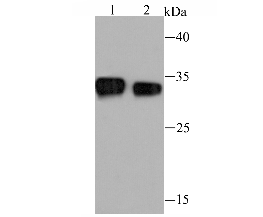
Fig1: Western blot analysis of ESD on K562 cell (1) and mouse intestine tissue (2) lysate using anti-ESD antibody at 1/1,000 dilution.Application
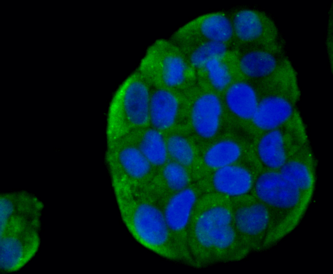
Fig2: ICC staining ESD in Hela cells (green). The nuclear counter stain is DAPI (blue). Cells were fixed in paraformaldehyde, permeabilised with 0.25% Triton X100/PBS.Application
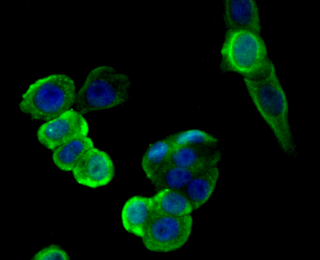
Fig3: ICC staining ESD in LOVO cells (green). The nuclear counter stain is DAPI (blue). Cells were fixed in paraformaldehyde, permeabilised with 0.25% Triton X100/PBS.Application
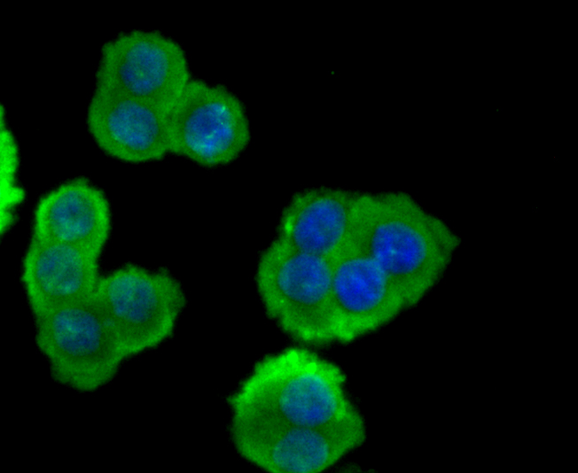
Fig4: ICC staining ESD in SW480 cells (green). The nuclear counter stain is DAPI (blue). Cells were fixed in paraformaldehyde, permeabilised with 0.25% Triton X100/PBS.Application
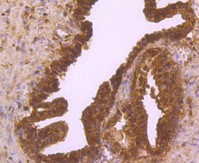
Fig5: Immunohistochemical analysis of paraffin-embedded human prostate tissue using anti-ESD antibody. Counter stained with hematoxylin.Application
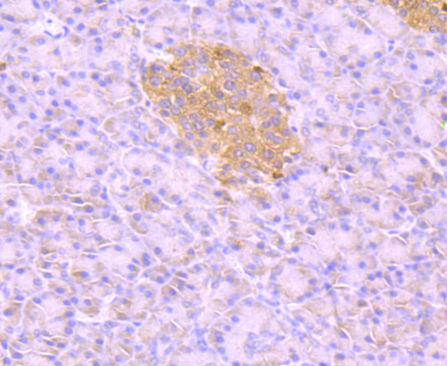
Fig6: Immunohistochemical analysis of paraffin-embedded human pancreas tissue using anti-ESD antibody. Counter stained with hematoxylin.Application
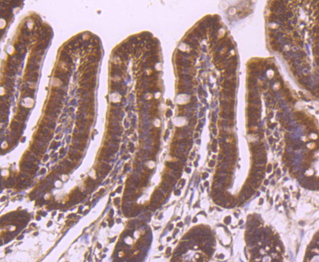
Fig7: Immunohistochemical analysis of paraffin-embedded mouse colon tissue using anti-e=ESD antibody. Counter stained with hematoxylin.| Product Name | ESD |
|---|---|
| Antibody Type | Primary Antibodies |
| Antigen Alias | EC 3.1.2.12 antibody Es-10 antibody Es10 antibody ESD antibody ESTD_HUMAN antibody Esterase 10 antibody Esterase D antibody Esterase D formylglutathione hydrolase antibody FGH antibody FGH, included antibody FLJ11763 antibody Methylumbelliferyl acetate deacetylase antibody MGC139609 antibody OTTHUMP00000018374 antibody OTTHUMP00000040929 antibody S formylglutathione hydrolase antibody S-formylglutathione hydrolase antibody S-formylglutathione hydrolase, included antibody Sid 478 antibody sid478 antibody |
| Product description | ESD (esterase D) is also known as S-formylglutathione hydrolase and is a 282 amino acid protein that is a member of the esterase D family. ESD is highly expressed in placenta, kidney, liver and erythrocytes, and is localized to the cytoplasm, as well as to cytoplasmic vesicles. The main function of ESD is to detoxify formaldehyde while providing energy. Formaldehyde is oxidized by ADH5 which yields S-formylglutathione. ESD then catalyzes the hydrolysis of S-formylglutathione to the reduced forms of formic acid and glutathione. In addition, ESD hydrolyzes a variety of different neutral ester substrates and can act as a carboxylesterase. ESD may also act as a cysteine hydrolase which is inactivated by thiol alkylating agents. ESD gene polymorphism can lead to reduced enzymatic activity which may cause susceptibility to many conditions, including toxic liver cirrhosis, retinoblastoma, obesity and autism. |
| Immunogen | Recombinant protein |
| Clonality | Polyclonal |
|---|---|
| Isotype | IgG |
| Host Species | Rabbit |
| Tested Applications | WBICCIHC |
| WB:1:500~1:2000 ICC:1:200~1:500 IHC:1:50~1:200 Notes:Optimal dilutions/concentrations should be determined by the researcher. |
|
| Species Reactivity | HumanMouse |
| Concentration | 1mg/ml |
| Alternative Names | EC 3.1.2.12 antibody
Es-10 antibody
Es10 antibody
ESD antibody
ESTD_HUMAN antibody
Esterase 10 antibody
Esterase D antibody
Esterase D formylglutathione hydrolase antibody
FGH antibody
FGH included antibody FLJ11763 antibody Methylumbelliferyl acetate deacetylase antibody MGC139609 antibody OTTHUMP00000018374 antibody OTTHUMP00000040929 antibody S formylglutathione hydrolase antibody S-formylglutathione hydrolase antibody S-formylglutathione hydrolase included antibody Sid 478 antibody sid478 antibody |
|---|---|
| Molecular Weight(MW) | 31 kDa |
| Cellular Localization | Cytoplasm. Cytoplasmic vesicle. |
Application
Fig1: Western blot analysis of ESD on K562 cell (1) and mouse intestine tissue (2) lysate using anti-ESD antibody at 1/1,000 dilution.
Application
Fig2: ICC staining ESD in Hela cells (green). The nuclear counter stain is DAPI (blue). Cells were fixed in paraformaldehyde, permeabilised with 0.25% Triton X100/PBS.
Application
Fig3: ICC staining ESD in LOVO cells (green). The nuclear counter stain is DAPI (blue). Cells were fixed in paraformaldehyde, permeabilised with 0.25% Triton X100/PBS.
Application
Fig4: ICC staining ESD in SW480 cells (green). The nuclear counter stain is DAPI (blue). Cells were fixed in paraformaldehyde, permeabilised with 0.25% Triton X100/PBS.
Application
Fig5: Immunohistochemical analysis of paraffin-embedded human prostate tissue using anti-ESD antibody. Counter stained with hematoxylin.
Application
Fig6: Immunohistochemical analysis of paraffin-embedded human pancreas tissue using anti-ESD antibody. Counter stained with hematoxylin.
Application
Fig7: Immunohistochemical analysis of paraffin-embedded mouse colon tissue using anti-e=ESD antibody. Counter stained with hematoxylin.| Positive Control | K562, mouse intestine tissue lysate, Hela, LOVO, SW480, human prostate tissue, human pancreas tissue, mouse colon tissue. |
|---|---|
| Application Notes | WB:1:500~1:2000 ICC:1:200~1:500 IHC:1:50~1:200 Notes:Optimal dilutions/concentrations should be determined by the researcher. |
| Form | Liquid |
|---|---|
| Storage Instructions | Store at +4℃after thawing. Aliquot store at -20℃or -80℃. Avoid repeated freeze / thaw cycles. |
| Storage Buffer | 1*TBS (pH7.4), 1%BSA, 40%Glycerol. Preservative: 0.05% Sodium Azide. |


