Application
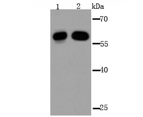
Fig1: Western blot analysis of IMP-3 on Hela (1) and 293T (2) cell lysate using anti-IMP-3 antibody at 1/2,000 dilution.Application
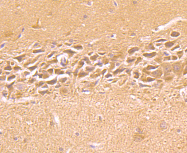
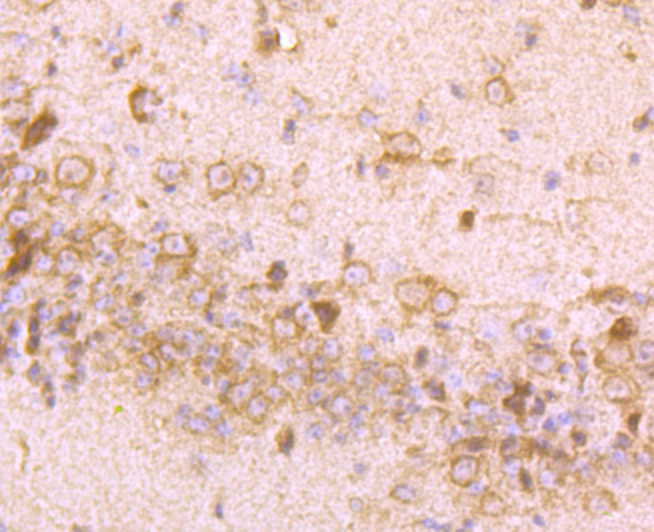
Fig2: Immunohistochemical analysis of paraffin-embedded rat brain tissue using anti-IMP-3 antibody. Counter stained with hematoxylin.Application
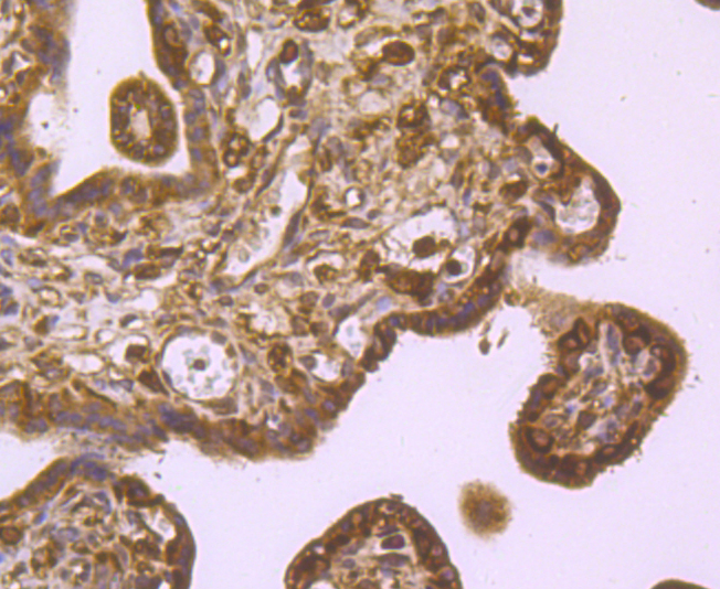
Fig3: Immunohistochemical analysis of paraffin-embedded human placental tissue using anti-IMP-3 antibody. Counter stained with hematoxylin.Application
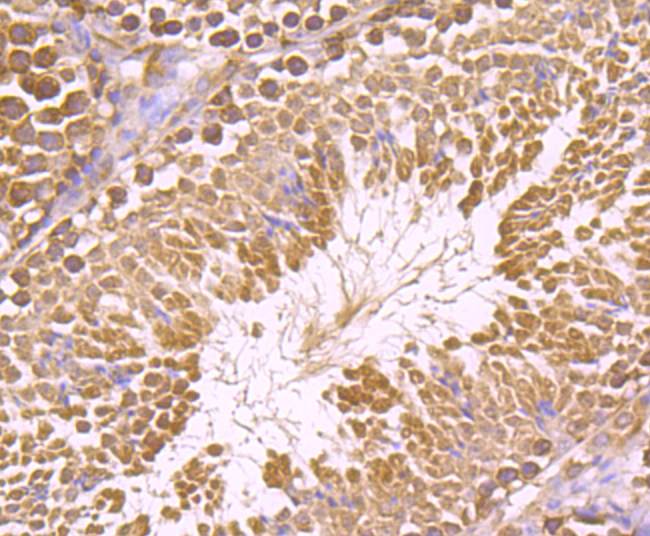
Fig4: Immunohistochemical analysis of paraffin-embedded mouse testis tissue using anti-IMP-3 antibody. Counter stained with hematoxylin.Application
Fig5: Immunohistochemical analysis of paraffin-embedded mouse brain tissue using anti-IMP-3 antibody. Counter stained with hematoxylin.Application
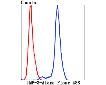
Fig6: Flow cytometric analysis of HepG2 cells with IPM-3 antibody at 1/100 dilution (red) compared with an unlabelled control (cells without incubation with primary antibody; black).| Product Name | IMP-3 |
|---|---|
| Antibody Type | Primary Antibodies |
| Antigen Alias | Cancer/testis antigen 98 antibody CT98 antibody DKFZp686F1078 antibody hKOC antibody IF2B3_HUMAN antibody IGF II mRNA binding protein 3 antibody IGF-II mRNA-binding protein 3 antibody IGF2 mRNA binding protein 3 antibody IGF2 mRNA-binding protein 3 antibody IGF2BP3 antibody IMP 3 antibody IMP-3 antibody Insulin like growth factor 2 mRNA binding protein 3 antibody Insulin-like growth factor 2 mRNA-binding protein 3 antibody KH domain containing protein overexpressed in cancer antibody KH domain-containing protein overexpressed in cancer antibody KOC 1 antibody KOC1 antibody VICKZ 3 antibody VICKZ family member 3 antibody VICKZ3 antibody |
| Product description | IGF-II mRNA-binding proteins (IMPs) bind RNA and influence RNA synthesis and metabolism. IMP-1, also known as coding region determinant-binding protein/insulin-like growth factor II mRNA-binding protein (CRD-BP) and VICKZ1; IMP-2 (IMP2, VICKZ2, p62); and IMP-3 (KOC1, VICKZ3) contain a unique combination of RNA recognition motifs and four hnRNP K homology domains. IMP-1 is abundant in embryonal tissues and is expressed in 81% of colon cancers, 73% of sarcomas and 58.5% of breast cancers. It recognizes c-Myc, IGF-II and t mRNAs, and H19 RNA, and plays a major role in proliferation of K-562 cells by an IGF-II-dependent mechanism. IMP-2 binds the 5' UTR of IGF-II mRNA and influences tumor cell growth, in which IMP-2 is associated with apoptosis induced by tretinoin. IMP-3 knockdown by RNA interference decreases levels of IGF-II protein without affecting IGF-II, c-Myc, or β Actin mRNA and H19 RNA levels. IMP-3 is a marker for carcinomas and high-grade dysplastic lesions of pancreatic ductal epithelium. |
| Immunogen | Recombinant protein |
| Clonality | Polyclonal |
|---|---|
| Isotype | IgG |
| Host Species | Rabbit |
| Tested Applications | WBIHCFC |
| WB:1:1,000-1:2,000 IHC:1:50-1 200FC:1:50-1:100 |
|
| Species Reactivity | HumanMouseRat |
| Concentration | 1mg/ml |
| Alternative Names | Cancer/testis antigen 98 antibody CT98 antibody DKFZp686F1078 antibody hKOC antibody IF2B3_HUMAN antibody IGF II mRNA binding protein 3 antibody IGF-II mRNA-binding protein 3 antibody IGF2 mRNA binding protein 3 antibody IGF2 mRNA-binding protein 3 antibody IGF2BP3 antibody IMP 3 antibody IMP-3 antibody Insulin like growth factor 2 mRNA binding protein 3 antibody Insulin-like growth factor 2 mRNA-binding protein 3 antibody KH domain containing protein overexpressed in cancer antibody KH domain-containing protein overexpressed in cancer antibody KOC 1 antibody KOC1 antibody VICKZ 3 antibody VICKZ family member 3 antibody VICKZ3 antibody |
|---|---|
| Molecular Weight(MW) | 63 kDa |
| Cellular Localization | Nucleus. Cytoplasm. |
Application
Fig1: Western blot analysis of IMP-3 on Hela (1) and 293T (2) cell lysate using anti-IMP-3 antibody at 1/2,000 dilution.
Application
Fig2: Immunohistochemical analysis of paraffin-embedded rat brain tissue using anti-IMP-3 antibody. Counter stained with hematoxylin.
Application
Fig3: Immunohistochemical analysis of paraffin-embedded human placental tissue using anti-IMP-3 antibody. Counter stained with hematoxylin.
Application
Fig4: Immunohistochemical analysis of paraffin-embedded mouse testis tissue using anti-IMP-3 antibody. Counter stained with hematoxylin.
Application
Fig5: Immunohistochemical analysis of paraffin-embedded mouse brain tissue using anti-IMP-3 antibody. Counter stained with hematoxylin.
Application
Fig6: Flow cytometric analysis of HepG2 cells with IPM-3 antibody at 1/100 dilution (red) compared with an unlabelled control (cells without incubation with primary antibody; black).| Positive Control | Hela, 293T, rat brain tissue, human placental tissue, mouse testis tissue, mouse brain tissue,HepG2. |
|---|---|
| Application Notes | WB:1:1,000-1:2,000 IHC:1:50-1 200FC:1:50-1:100 |
| Form | Liquid |
|---|---|
| Storage Instructions | Store at +4℃ after thawing. Aliquot store at -20℃ or -80℃. Avoid repeated freeze / thaw cycles. |
| Storage Buffer | 1*TBS (pH7.4), 1%BSA, 40%Glycerol. Preservative: 0.05% Sodium Azide. |


