Application
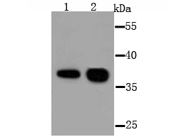
Fig1: Western blot analysis of AKR1C1 on different lysate using anti-AKR1C1 antibody at 1/1,000 dilution.Positive control:Lane1: Mouse testisLane2: SW480Application
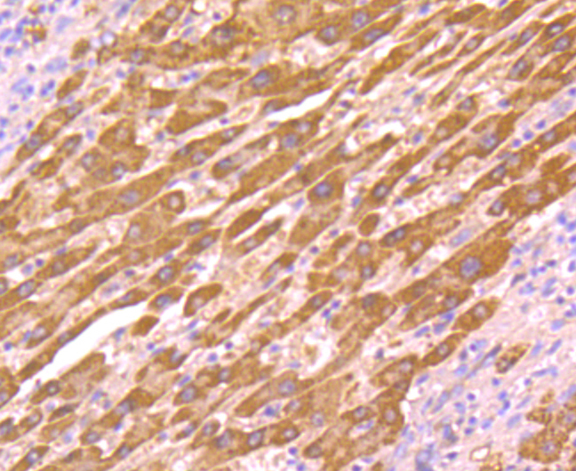
Fig2: Immunohistochemical analysis of paraffin-embedded human liver cancer tissue using anti-AKR1C1 antibody. Counter stained with hematoxylin.Application
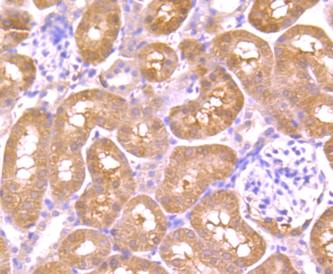
Fig3: Immunohistochemical analysis of paraffin-embedded mouse kidney tissue using anti-AKR1C1 antibody. Counter stained with hematoxylin.Application
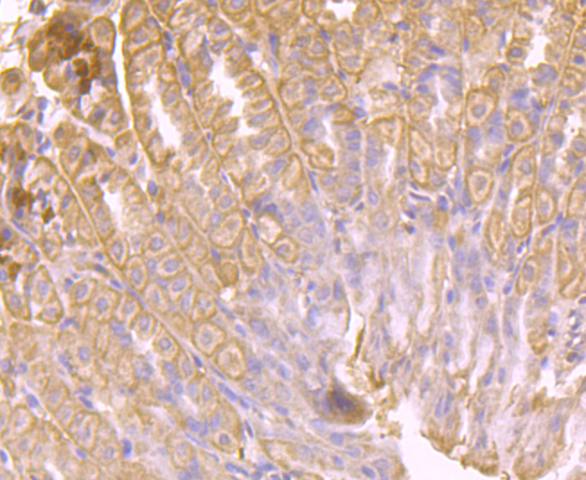
Fig4: Immunohistochemical analysis of paraffin-embedded mouse stomach tissue using anti-AKR1C1 antibody. Counter stained with hematoxylin.Application
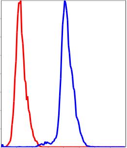
Fig5: Flow cytometric analysis of HepG2 cells with AKR1C1 antibody at 1/100 dilution (blue) compared with an unlabelled control (cells without incubation with primary antibody; red).| Product Name | AKR1C1 |
|---|---|
| Antibody Type | Primary Antibodies |
| Antigen Alias | 2-dihydrobenzene-1 antibody 2-diol dehydrogenase antibody 20 alpha (3 alpha) hydroxysteroid dehydrogenase antibody 20 ALPHA HSD antibody 20 alpha hydroxysteroid dehydrogenase antibody 20-alpha-HSD antibody 20-alpha-hydroxysteroid dehydrogenase antibody 20ALPHAHSD antibody 2ALPHAHSD antibody AK1C1 antibody AK1C1_HUMAN antibody AKR1C1 antibody Aldo keto reductase family 1 member C1 antibody aldo-keto reductase C antibody Aldo-keto reductase family 1 member C1 antibody Aldo-keto reductase family, 1 member 1 antibody C9 antibody Chlordecone reductase homolog antibody Chlordecone reductase homolog HAKRC antibody DD1 antibody DD1/DD2 antibody DDH antibody DDH1 antibody Dihydrodiol dehydrogenase 1 antibody Dihydrodiol dehydrogenase 1/2 antibody dihydrodiol dehydrogenase 1; 20-alpha (3-alpha)-hydroxysteroid dehydrogenase antibody dihydrodiol dehydrogenase isoform DD1 antibody Dihydrodiol dehydrogenase, type 1 antibody H37 antibody HAKRC antibody HBAB antibody Hepatic dihydrodiol dehydrogenase antibody High affinity hepatic bile acid-binding protein antibody High-affinity hepatic bile acid-binding protein antibody Indanol dehydrogenase antibody MBAB antibody MGC8954 antibody Trans-1 antibody Trans-1,2 dihydrobenzene 1,2 diol dehydrogenase antibody Type II 3 alpha hydroxysteroid dehydrogenase antibody |
| Product description | Human liver contains isoforms of dihydrodiol dehydrogenase (DD1, DD2, DD3 and DD4), which belong to the aldo-oxo reductase/aldo-keto reductase (AKR) superfamily, have 20α- or 3α-hydroxysteroid dehydrogenase (HSD) activity. DD1 is also designated AKR1C1, DDH or DDH1, while DD2 also can be designated AKR1C2, dDD, BABP or DDH2. AKR1C3 and 3α-HSD are alternate designations for human DD3 (which is referred to as AKR1C18 in rodents), while DD4 also can be called AKR1C4, CD, CHDR or AKR1C6 (in rodents). DD1 and DD2 are 20α-HSDs, whereas DD3 and DD4 are the 3α-HSDs. The multiple human cytosolic dihydrodiol dehydrogenases are involved in the metabolism of xenobiotics, such as polycyclic aromatic hydrocarbons, pesticides and steroid hormones, and are responsible for the reduction of ketone-containing drugs by using NADH or NADPH as a cofactor. The 20α-HSD catalyzes the reaction of progesterone to the inactive form 20α-hydroxyprogesterone. The 3α-HSD is a cytosolic, monomeric, NADPH-dependent oxidoreductase that reduces 3-keto-5-dihydrosteroids to their tetrahydro products. DD1 and DD2 are ubiquitously expressed, whereas DD4 mRNA is restricted to the liver. DD3 is a unique enzyme that can specifically catalyze the dehydrogenation of trans-benzenedihydrodiol and trans-naphthalenedihydrodiol. |
| Immunogen | recombinant protein |
| Clonality | Polyclonal |
|---|---|
| Isotype | IgG |
| Host Species | Rabbit |
| Tested Applications | WBICCIHCFC |
| WB:1:500~1:2000 ICC:1:200~1:500 IHC:1:50~1:200 FC:1:200~1:500 Notes:Optimal dilutions/concentrations should be determined by the researcher. |
|
| Species Reactivity | HumanMouse |
| Concentration | 1mg/ml |
| Alternative Names | 2-dihydrobenzene-1 antibody
2-diol dehydrogenase antibody
20 alpha (3 alpha) hydroxysteroid dehydrogenase antibody
20 ALPHA HSD antibody
20 alpha hydroxysteroid dehydrogenase antibody
20-alpha-HSD antibody
20-alpha-hydroxysteroid dehydrogenase antibody
20ALPHAHSD antibody
2ALPHAHSD antibody
AK1C1 antibody
AK1C1_HUMAN antibody
AKR1C1 antibody
Aldo keto reductase family 1 member C1 antibody
aldo-keto reductase C antibody
Aldo-keto reductase family 1 member C1 antibody
Aldo-keto reductase family 1 member 1 antibody C9 antibody Chlordecone reductase homolog antibody Chlordecone reductase homolog HAKRC antibody DD1 antibody DD1/DD2 antibody DDH antibody DDH1 antibody Dihydrodiol dehydrogenase 1 antibody Dihydrodiol dehydrogenase 1/2 antibody dihydrodiol dehydrogenase 1 20-alpha (3-alpha)-hydroxysteroid dehydrogenase antibody dihydrodiol dehydrogenase isoform DD1 antibody Dihydrodiol dehydrogenase type 1 antibody H37 antibody HAKRC antibody HBAB antibody Hepatic dihydrodiol dehydrogenase antibody High affinity hepatic bile acid-binding protein antibody High-affinity hepatic bile acid-binding protein antibody Indanol dehydrogenase antibody MBAB antibody MGC8954 antibody Trans-1 antibody Trans-1 2 dihydrobenzene 1 2 diol dehydrogenase antibody Type II 3 alpha hydroxysteroid dehydrogenase antibody |
|---|---|
| Molecular Weight(MW) | 37 kDa |
| Cellular Localization | Cytoplasm. |
| SwissProt ID | Q04828 |
|---|
Application
Fig1: Western blot analysis of AKR1C1 on different lysate using anti-AKR1C1 antibody at 1/1,000 dilution.Positive control:Lane1: Mouse testisLane2: SW480
Application
Fig2: Immunohistochemical analysis of paraffin-embedded human liver cancer tissue using anti-AKR1C1 antibody. Counter stained with hematoxylin.
Application
Fig3: Immunohistochemical analysis of paraffin-embedded mouse kidney tissue using anti-AKR1C1 antibody. Counter stained with hematoxylin.
Application
Fig4: Immunohistochemical analysis of paraffin-embedded mouse stomach tissue using anti-AKR1C1 antibody. Counter stained with hematoxylin.
Application
Fig5: Flow cytometric analysis of HepG2 cells with AKR1C1 antibody at 1/100 dilution (blue) compared with an unlabelled control (cells without incubation with primary antibody; red).| Positive Control | Mouse testes tissue lysate, SW1990 cell lysate, human liver cancer tissue, mouse kidney tissue, mouse stomach tissue, HepG2. |
|---|---|
| Application Notes | WB:1:500~1:2000 ICC:1:200~1:500 IHC:1:50~1:200 FC:1:200~1:500 Notes:Optimal dilutions/concentrations should be determined by the researcher. |
| Form | Liquid |
|---|---|
| Storage Instructions | Store at +4℃ after thawing. Aliquot store at -20℃ or -80℃. Avoid repeated freeze / thaw cycles. |
| Storage Buffer | 1*TBS (pH7.4), 1%BSA, 40%Glycerol. Preservative: 0.05% Sodium Azide. |


