Application
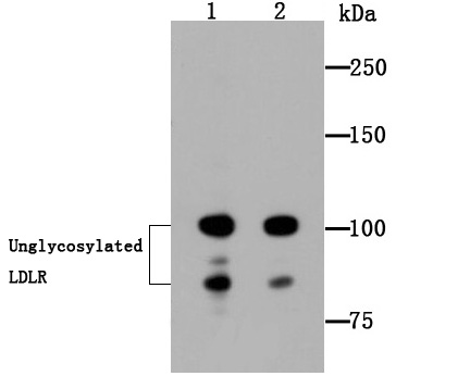
Fig1: Western blot analysis of LDL Receptor on Hela (1) and MCF-7 (2) cell lysates using anti-LDL Receptor antibody at 1/1,000 dilution.Application
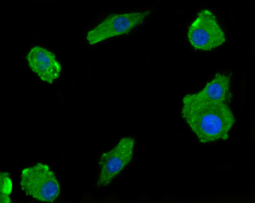
Fig2: ICC staining LDL Receptor in A549 cells (red). The nuclear counter stain is DAPI (blue). Cells were fixed in paraformaldehyde, permeabilised with 0.25% Triton X100/PBS.Application
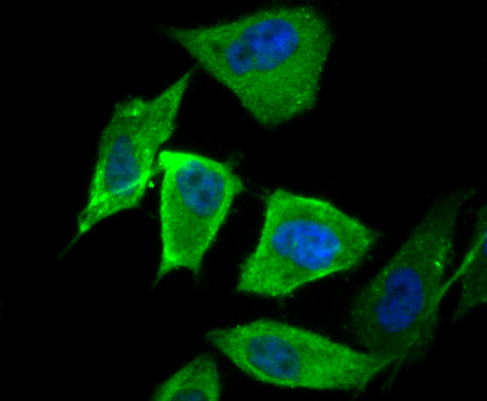
Fig3: ICC staining LDL Receptor in PC-3M cells (red). The nuclear counter stain is DAPI (blue). Cells were fixed in paraformaldehyde, permeabilised with 0.25% Triton X100/PBS.Application
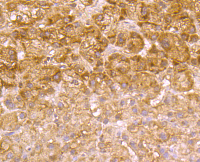
Fig4: Immunohistochemical analysis of paraffin-embedded human liver cancer tissue using anti-LDL Receptor antibody. Counter stained with hematoxylin.Application
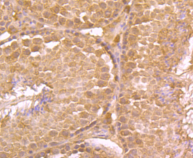
Fig5: Immunohistochemical analysis of paraffin-embedded mouse testis tissue using anti-LDL Receptor antibody. Counter stained with hematoxylin.Application
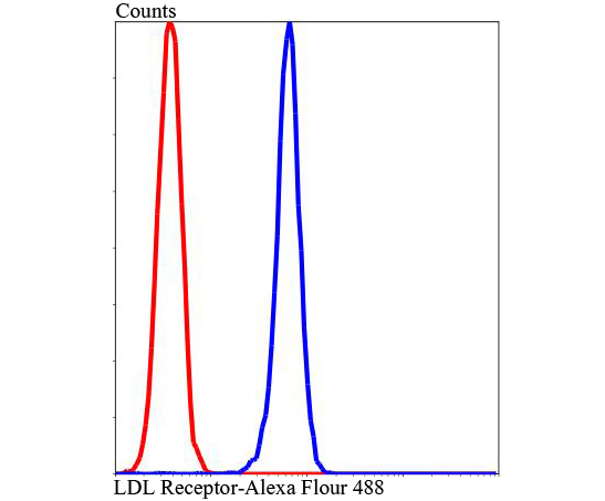
Fig6: Flow cytometric analysis of Hela cells with LDL Receptor antibody at 1/100 dilution (blue) compared with an unlabelled control (cells without incubation with primary antibody; red). Alexa Fluor 488-conjugated Goat anti rabbit IgG was used as the secondary antibody.| Product Name | LDL Receptor |
|---|---|
| Antibody Type | Primary Antibodies |
| Antigen Alias | FH antibody FHC antibody LDL R antibody LDL receptor antibody LDLCQ2 antibody Ldlr antibody LDLR_HUMAN antibody Low Density Lipoprotein Receptor antibody Low density lipoprotein receptor class A domain containing protein 3 antibody Low density lipoprotein receptor familial hypercholesterolemia antibody Low-density lipoprotein receptor antibody |
| Product description | LDLR (low density lipoprotein receptor) is a member of the LDL receptor gene family, which includes LDLR, LRP, Megalin, VLDLR, and ApoER2. The LDL receptor family is characterized by a cluster of cysteine-rich class A repeats, epidermal growth factor (EGF)-like repeats, YWTD repeats, and an O-linked sugar domain. The LDL receptor is a cell surface transmembrane protein that mediates the uptake of low density lipoprotein and its degradation in the lysosome, which provides cholesterol to cells. The cytoplasmic domain of the LDL receptor is necessary for the receptor to cluster in coated pits, which promotes the rapid endocytosis of bound LDL. Mutations in LDLR cause the autosomal dominant disease, familial hypercholesterolemia (FH), which promotes premature coronary atherosclerosis. |
| Immunogen | Peptide. |
| Clonality | Polyclonal |
|---|---|
| Isotype | IgG |
| Host Species | Rabbit |
| Tested Applications | WBICCIHCFC |
| WB:1:1,000-1:5,000 ICC:1:50-1:200 IHC:1:50-1:200 FC:1:50-1:100 |
|
| Species Reactivity | HumanMouse |
| Concentration | 1mg/ml |
| Alternative Names | FH antibody FHC antibody LDL R antibody LDL receptor antibody LDLCQ2 antibody Ldlr antibody LDLR_HUMAN antibody Low Density Lipoprotein Receptor antibody Low density lipoprotein receptor class A domain containing protein 3 antibody Low density lipoprotein receptor familial hypercholesterolemia antibody Low-density lipoprotein receptor antibody |
|---|---|
| Molecular Weight(MW) | 95/76 kDa(Predicted band size) |
| Cellular Localization | Golgi apparatus. Plasma membrane. Endosome. Lysosome. |
Application
Fig1: Western blot analysis of LDL Receptor on Hela (1) and MCF-7 (2) cell lysates using anti-LDL Receptor antibody at 1/1,000 dilution.
Application
Fig2: ICC staining LDL Receptor in A549 cells (red). The nuclear counter stain is DAPI (blue). Cells were fixed in paraformaldehyde, permeabilised with 0.25% Triton X100/PBS.
Application
Fig3: ICC staining LDL Receptor in PC-3M cells (red). The nuclear counter stain is DAPI (blue). Cells were fixed in paraformaldehyde, permeabilised with 0.25% Triton X100/PBS.
Application
Fig4: Immunohistochemical analysis of paraffin-embedded human liver cancer tissue using anti-LDL Receptor antibody. Counter stained with hematoxylin.
Application
Fig5: Immunohistochemical analysis of paraffin-embedded mouse testis tissue using anti-LDL Receptor antibody. Counter stained with hematoxylin.
Application
Fig6: Flow cytometric analysis of Hela cells with LDL Receptor antibody at 1/100 dilution (blue) compared with an unlabelled control (cells without incubation with primary antibody; red). Alexa Fluor 488-conjugated Goat anti rabbit IgG was used as the secondary antibody.| Positive Control | Hela, MCF-7,A541, PC-3M, human liver cancer tissue, mouse testis tissue. |
|---|---|
| Application Notes | WB:1:1,000-1:5,000 ICC:1:50-1:200 IHC:1:50-1:200 FC:1:50-1:100 |
| Form | Liquid |
|---|---|
| Storage Instructions | Store at +4℃ after thawing. Aliquot store at -20℃ or -80℃. Avoid repeated freeze / thaw cycles. |
| Storage Buffer | 1*TBS (pH7.4), 0.5%BSA, 50%Glycerol. Preservative: 0.05% Sodium Azide. |


