Application
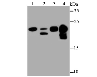
Fig1: Western blot analysis of Myosin Light Chain 2 on different lysates using anti-Myosin Light Chain 2 antibody at 1/500 dilution. Positive control: Lane1: Mouse skeletal muscle Lane2: Human skeletal muscle Lane3: Mouse heart Lane4: Rat heartApplication
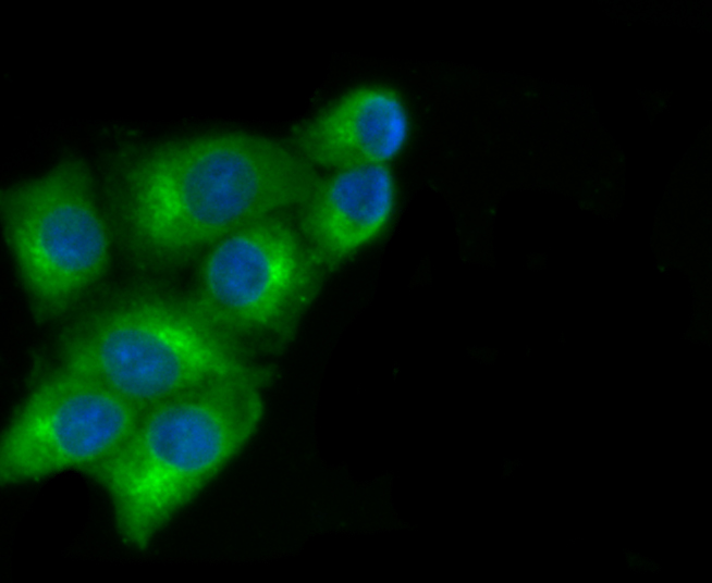
Fig2: ICC staining Myosin Light Chain 2 in A549 cells (green). The nuclear counter stain is DAPI (blue). Cells were fixed in paraformaldehyde, permeabilised with 0.25% Triton X100/PBS.Application
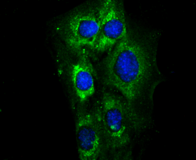
Fig3: ICC staining Myosin Light Chain 2 in C2C12 cells (green). The nuclear counter stain is DAPI (blue). Cells were fixed in paraformaldehyde, permeabilised with 0.25% Triton X100/PBS.Application
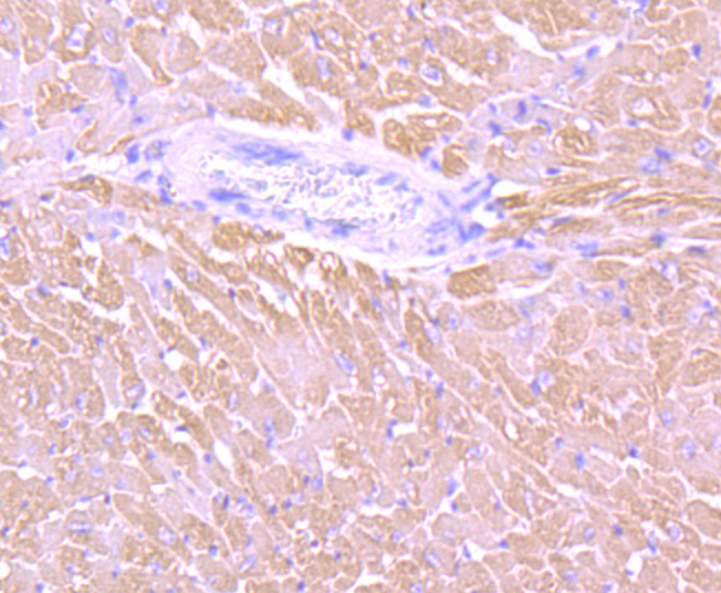
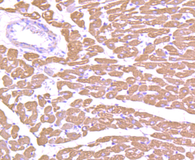
Fig4: Immunohistochemical analysis of paraffin-embedded mouse heart tissue using anti-Myosin Light Chain 2 antibody. Counter stained with hematoxylin.Application
Fig5: Immunohistochemical analysis of paraffin-embedded rat heart tissue using anti-Myosin Light Chain 2 antibody. Counter stained with hematoxylin.Application
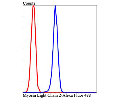
Fig6: Flow cytometric analysis of Hela cells with Myosin Light Chain 2 antibody at 1/100 dilution (blue) compared with an unlabelled control (cells without incubation with primary antibody; red). Alexa Fluor 488-conjugated Goat anti rabbit IgG was used as the secondary antibody.| Product Name | Myosin Light Chain 2 |
|---|---|
| Antibody Type | Primary Antibodies |
| Antigen Alias | Cardiac myosin light chain-2 antibody Cardiac ventricular myosin light chain 2 antibody CMH10 antibody MLC 2v antibody MLC-2 antibody MLC-2v antibody MLC2 antibody MLRV_HUMAN antibody MYL 2 antibody MYL2 antibody Myosin light chain 2 regulatory cardiac slow antibody Myosin light polypeptide 2 regulatory cardiac slow antibody Myosin regulatory light chain 2 antibody Myosin regulatory light chain 2 ventricular/cardiac muscle isoform antibody Regulatory light chain of myosin antibody RLC of myosin antibody Slow cardiac myosin regulatory light chain 2 antibody ventricular/cardiac muscle isoform antibody |
| Product description | Encoded by the MYL2 gene, myosin regulatory light chain 2, ventricular/cardiac muscle isoform, also designated MLC-2 or MLC2v, is part of a hexameric complex of two heavy chains and four light chains predominantly expressed in adult cardiac ventricle muscle. Myosin regulatory light chain 2 binds calcium and has been shown to be a useful molecular marker for cardiac chamber specification. The co-expression of myosin regulatory light chain 7 (MYL7) and myosin regulatory light chain 2 in the outflow tract and atrioventricular canal, together with the single expression in the atrial (MYL7) and ventricular (MYL2) myocardium, permits the delineation of their boundaries. At the amino acid level there is 96% homology between the human and mouse myosin regulatory light chain sequences. Mutations in MYL2 are correlated with mid-left ventricular chamber type hypertrophic cardiomyopathy (MVC2) as well as familial hypertrophic cardiomyopathy type 10 (CMH10). |
| Immunogen | Peptide |
| Clonality | Polyclonal |
|---|---|
| Isotype | IgG |
| Host Species | Rabbit |
| Tested Applications | WBICCIHCFC |
| WB:1:1,000-1:2,000 ICC:1:50-1:200 IHC:1:50-1:200 FC:1:50-1:100 |
|
| Species Reactivity | HumanMouseRat |
| Concentration | 1mg/ml |
| Alternative Names | Cardiac myosin light chain-2 antibody Cardiac ventricular myosin light chain 2 antibody CMH10 antibody MLC 2v antibody MLC-2 antibody MLC-2v antibody MLC2 antibody MLRV_HUMAN antibody MYL 2 antibody MYL2 antibody Myosin light chain 2 regulatory cardiac slow antibody Myosin light polypeptide 2 regulatory cardiac slow antibody Myosin regulatory light chain 2 antibody Myosin regulatory light chain 2 ventricular/cardiac muscle isoform antibody Regulatory light chain of myosin antibody RLC of myosin antibody Slow cardiac myosin regulatory light chain 2 antibody ventricular/cardiac muscle isoform antibody |
|---|---|
| Molecular Weight(MW) | 19 kDa |
| Cellular Localization | Cytoplasm. |
Application
Fig1: Western blot analysis of Myosin Light Chain 2 on different lysates using anti-Myosin Light Chain 2 antibody at 1/500 dilution. Positive control: Lane1: Mouse skeletal muscle Lane2: Human skeletal muscle Lane3: Mouse heart Lane4: Rat heart
Application
Fig2: ICC staining Myosin Light Chain 2 in A549 cells (green). The nuclear counter stain is DAPI (blue). Cells were fixed in paraformaldehyde, permeabilised with 0.25% Triton X100/PBS.
Application
Fig3: ICC staining Myosin Light Chain 2 in C2C12 cells (green). The nuclear counter stain is DAPI (blue). Cells were fixed in paraformaldehyde, permeabilised with 0.25% Triton X100/PBS.
Application
Fig4: Immunohistochemical analysis of paraffin-embedded mouse heart tissue using anti-Myosin Light Chain 2 antibody. Counter stained with hematoxylin.
Application
Fig5: Immunohistochemical analysis of paraffin-embedded rat heart tissue using anti-Myosin Light Chain 2 antibody. Counter stained with hematoxylin.
Application
Fig6: Flow cytometric analysis of Hela cells with Myosin Light Chain 2 antibody at 1/100 dilution (blue) compared with an unlabelled control (cells without incubation with primary antibody; red). Alexa Fluor 488-conjugated Goat anti rabbit IgG was used as the secondary antibody.| Positive Control | Mouse skeletal muscle tissue lysate, human skeletal muscle tissue lysate, mouse heart tissue, rat heart tissue, A549, Hela, C2C12. |
|---|---|
| Application Notes | WB:1:1,000-1:2,000 ICC:1:50-1:200 IHC:1:50-1:200 FC:1:50-1:100 |
| Form | Liquid |
|---|---|
| Storage Instructions | Store at +4℃ after thawing. Aliquot store at -20℃ or -80℃. Avoid repeated freeze / thaw cycles. |
| Storage Buffer | 1*TBS (pH7.4), 0.5%BSA, 50%Glycerol. Preservative: 0.05% Sodium Azide. |


