Application
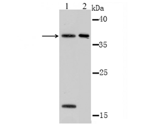
Fig1: Western blot analysis of OGG1 on F9 cell(1) and mouse kidney tissue (2) lysate using anti-OGG1 antibody at 1/1,000 dilution.Application
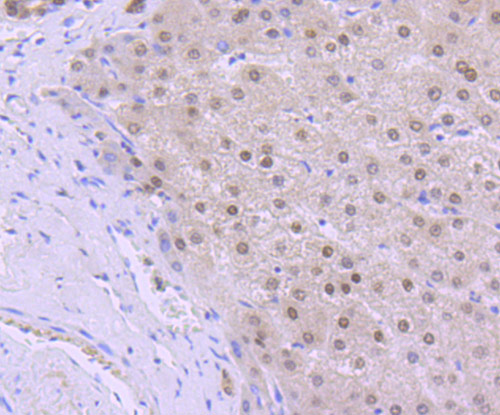
Fig2: Immunohistochemical analysis of paraffin-embedded human liver tissue using anti-OGG1 antibody. Counter stained with hematoxylin.Application
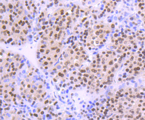
Fig3: Immunohistochemical analysis of paraffin-embedded human liver cancer tissue using anti-OGG1 antibody. Counter stained with hematoxylin.Application
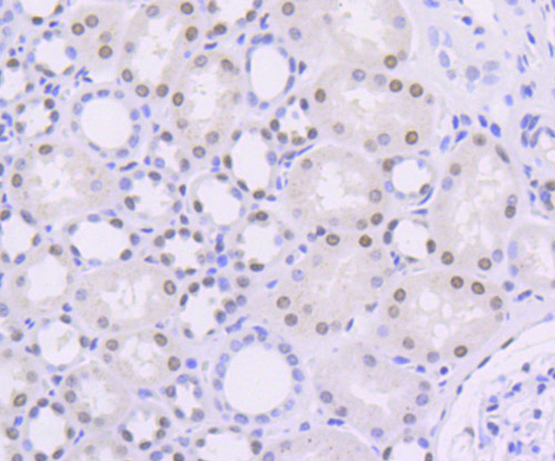
Fig4: Immunohistochemical analysis of paraffin-embedded human kidney tissue using anti-OGG1 antibody. Counter stained with hematoxylin.| Product Name | OGG1 |
|---|---|
| Antibody Type | Primary Antibodies |
| Antigen Alias | 8 hydroxyguanine DNA glycosylase antibody 8 oxoguanine DNA glycosylase 1 antibody 8 oxoguanine DNA glycosylase antibody AP lyase antibody DNA (apurinic or apyrimidinic site) lyase antibody DNA apurinic or apyrimidinic site lyase antibody DNA lyase antibody DNA-(apurinic or apyrimidinic site) lyase antibody HMMH antibody HOGG 1 antibody HOGG1 antibody MMH antibody MUTM antibody N glycosylase antibody Ogg 1 antibody Ogg1 antibody OGG1_HUMAN antibody OGH 1 antibody OGH1 antibody |
| Product description | DNA repair enzyme that incises DNA at 8-oxoG residues. Excises 7,8-dihydro-8-oxoguanine and 2,6-diamino-4-hydroxy-5-N-methylformamidopyrimidine (FAPY) from damaged DNA. Has a beta-lyase activity that nicks DNA 3' to the lesion. Defects in OGG1 may be a cause of renal cell carcinoma (RCC). It is a heterogeneous group of sporadic or hereditary carcinoma derived from cells of the proximal renal tubular epithelium. It is subclassified into clear cell renal carcinoma (non-papillary carcinoma), papillary renal cell carcinoma, chromophobe renal cell carcinoma, collecting duct carcinoma with medullary carcinoma of the kidney, and unclassified renal cell carcinoma. |
| Immunogen | Recombinant protein. |
| Clonality | Polyclonal |
|---|---|
| Isotype | IgG |
| Host Species | Rabbit |
| Tested Applications | WBIHC |
| WB:1:500~1:2000 IHC:1:50~1:200 Notes:Optimal dilutions/concentrations should be determined by the researcher. |
|
| Species Reactivity | HumanMouse |
| Concentration | 1mg/ml |
| Alternative Names | 8 hydroxyguanine DNA glycosylase antibody 8 oxoguanine DNA glycosylase 1 antibody 8 oxoguanine DNA glycosylase antibody AP lyase antibody DNA (apurinic or apyrimidinic site) lyase antibody DNA apurinic or apyrimidinic site lyase antibody DNA lyase antibody DNA-(apurinic or apyrimidinic site) lyase antibody HMMH antibody HOGG 1 antibody HOGG1 antibody MMH antibody MUTM antibody N glycosylase antibody Ogg 1 antibody Ogg1 antibody OGG1_HUMAN antibody OGH 1 antibody OGH1 antibody |
|---|---|
| Molecular Weight(MW) | 39 kDa |
| Cellular Localization | Mitochondrion. Nucleus. |
Application
Fig1: Western blot analysis of OGG1 on F9 cell(1) and mouse kidney tissue (2) lysate using anti-OGG1 antibody at 1/1,000 dilution.
Application
Fig2: Immunohistochemical analysis of paraffin-embedded human liver tissue using anti-OGG1 antibody. Counter stained with hematoxylin.
Application
Fig3: Immunohistochemical analysis of paraffin-embedded human liver cancer tissue using anti-OGG1 antibody. Counter stained with hematoxylin.
Application
Fig4: Immunohistochemical analysis of paraffin-embedded human kidney tissue using anti-OGG1 antibody. Counter stained with hematoxylin.| Positive Control | F9, mouse kidney tissue lysate, human liver tissue, human liver cancer tissue, human kidney tissue. |
|---|---|
| Application Notes | WB:1:500~1:2000 IHC:1:50~1:200 Notes:Optimal dilutions/concentrations should be determined by the researcher. |
| Form | Liquid |
|---|---|
| Storage Instructions | Store at +4℃ after thawing. Aliquot store at -20℃ or -80℃. Avoid repeated freeze / thaw cycles. |
| Storage Buffer | 1*TBS (pH7.4), 0.5%BSA, 50%Glycerol. Preservative: 0.05% Sodium Azide. |


