Application
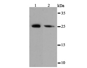
Fig1: Western blot analysis of eIF-4E on different cell lysates using anti-eIF-4E antibody at 1/500 dilution. Positive control: Lane1: PC-12 Lane2: NIH-3T3Application
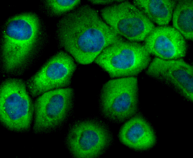
Fig2: ICC staining eIF-4E in A431 cells (green). The nuclear counter stain is DAPI (blue). Cells were fixed in paraformaldehyde, permeabilised with 0.25% Triton X100/PBS.Application
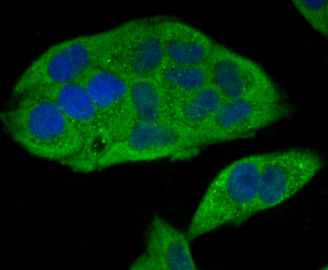
Fig3: ICC staining eIF-4E in Hela cells (green). The nuclear counter stain is DAPI (blue). Cells were fixed in paraformaldehyde, permeabilised with 0.25% Triton X100/PBS.Application
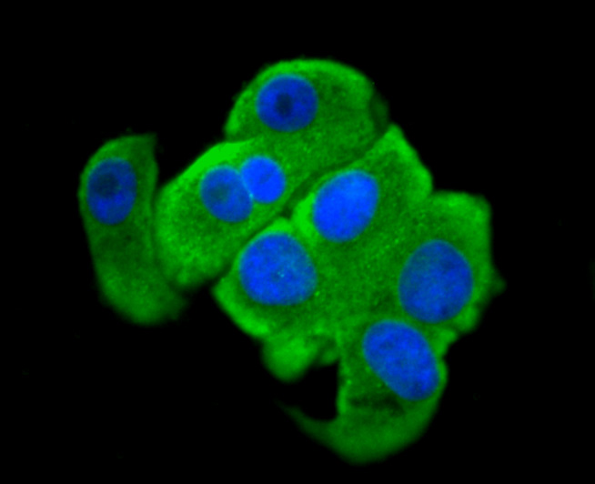
Fig4: ICC staining eIF-4E in SK-Br-3 cells (green). The nuclear counter stain is DAPI (blue). Cells were fixed in paraformaldehyde, permeabilised with 0.25% Triton X100/PBS.Application
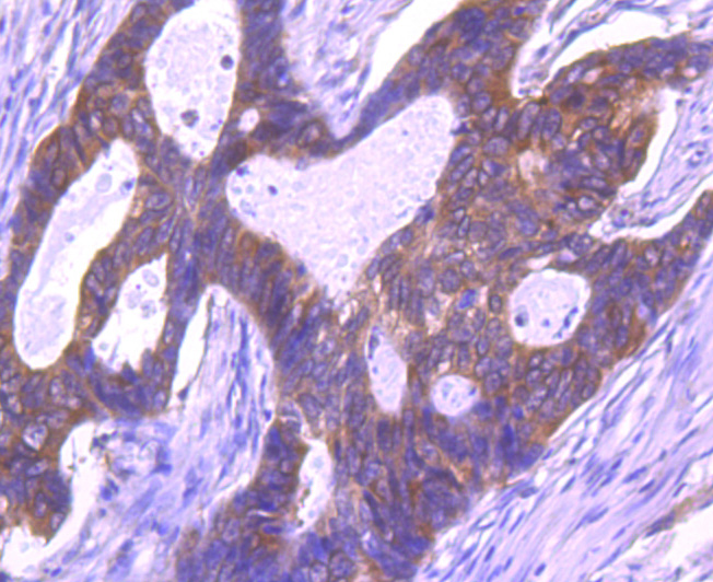
Fig5: Immunohistochemical analysis of paraffin-embedded human colon cancer tissue using anti-eIF-4E antibody. Counter stained with hematoxylin.Application
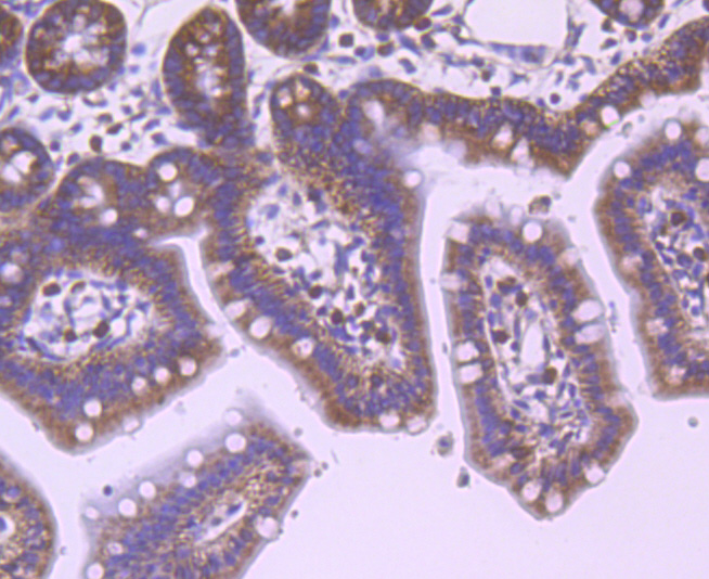
Fig6: Immunohistochemical analysis of paraffin-embedded mouse colon tissue using anti-eIF-4E antibody. Counter stained with hematoxylin.Application
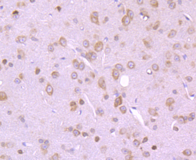
Fig7: Immunohistochemical analysis of paraffin-embedded mouse brain tissue using anti-eIF-4E antibody. Counter stained with hematoxylin.Application
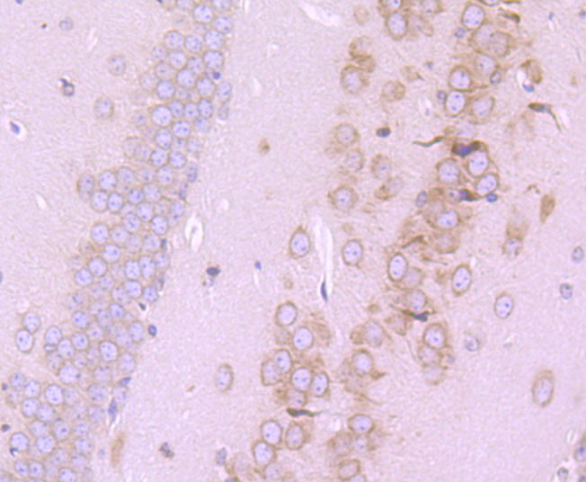
Fig8: Immunohistochemical analysis of paraffin-embedded mouse hippocampus tissue using anti-eIF-4E antibody. Counter stained with hematoxylin.Application
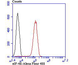
Fig9: Flow cytometric analysis of 293T cells with eIF-4E antibody at 1/100 dilution (red) compared with an unlabelled control (cells without incubation with primary antibody; black).| Product Name | eIF-4E |
|---|---|
| Antibody Type | Primary Antibodies |
| Antigen Alias | AUTS19 antibody CBP antibody eIF 4E antibody eIF 4F 25 kDa subunit antibody EIF 4F antibody eIF-4E antibody eIF-4F 25 kDa subunit antibody eIF4E antibody EIF4E1 antibody EIF4EL1 antibody EIF4F antibody Eukaryotic translation initiation factor 4 E antibody Eukaryotic translation initiation factor 4E antibody Eukaryotic translation initiation factor 4E like 1 antibody IF4E_HUMAN antibody Messanger RNA Cap Binding Protein eIF 4E antibody MGC111573 antibody mRNA cap binding protein antibody mRNA cap-binding protein antibody |
| Product description | The initiation of protein synthesis in eukaryotic cells is regulated by interactions between protein initiation factors and RNA molecules. The eukaryotic initiation complex eIF4F exists in vitro as a trimeric complex of eIF4E, eIF4A and eIF4G. Together, the complex allows ribosome binding to mRNA by inducing the unwinding of mRNA secondary structures. eIF4E binds to the mRNA "cap" during an early step in the initiation of protein synthesis. eIF4A acts as an ATP-dependent RNA helicase. eIF4G acts as a bridge between eIF4E, eIF4A and the eIF3 complex. |
| Immunogen | Peptide |
| Clonality | Polyclonal |
|---|---|
| Isotype | IgG |
| Host Species | Rabbit |
| Tested Applications | WBICCIHCFC |
| WB:1:500-1:1,000 ICC:1:50-1:200 IHC:1:50-1:100 FC:1:50-1:100 |
|
| Species Reactivity | HumanMouseRat |
| Concentration | 1mg/ml |
| Alternative Names | AUTS19 antibody CBP antibody eIF 4E antibody eIF 4F 25 kDa subunit antibody EIF 4F antibody eIF-4E antibody eIF-4F 25 kDa subunit antibody eIF4E antibody EIF4E1 antibody EIF4EL1 antibody EIF4F antibody Eukaryotic translation initiation factor 4 E antibody Eukaryotic translation initiation factor 4E antibody Eukaryotic translation initiation factor 4E like 1 antibody IF4E_HUMAN antibody Messanger RNA Cap Binding Protein eIF 4E antibody MGC111573 antibody mRNA cap binding protein antibody mRNA cap-binding protein antibody |
|---|---|
| Molecular Weight(MW) | 25 kDa |
| Cellular Localization | Cytoplasm. |
Application
Fig1: Western blot analysis of eIF-4E on different cell lysates using anti-eIF-4E antibody at 1/500 dilution. Positive control: Lane1: PC-12 Lane2: NIH-3T3
Application
Fig2: ICC staining eIF-4E in A431 cells (green). The nuclear counter stain is DAPI (blue). Cells were fixed in paraformaldehyde, permeabilised with 0.25% Triton X100/PBS.
Application
Fig3: ICC staining eIF-4E in Hela cells (green). The nuclear counter stain is DAPI (blue). Cells were fixed in paraformaldehyde, permeabilised with 0.25% Triton X100/PBS.
Application
Fig4: ICC staining eIF-4E in SK-Br-3 cells (green). The nuclear counter stain is DAPI (blue). Cells were fixed in paraformaldehyde, permeabilised with 0.25% Triton X100/PBS.
Application
Fig5: Immunohistochemical analysis of paraffin-embedded human colon cancer tissue using anti-eIF-4E antibody. Counter stained with hematoxylin.
Application
Fig6: Immunohistochemical analysis of paraffin-embedded mouse colon tissue using anti-eIF-4E antibody. Counter stained with hematoxylin.
Application
Fig7: Immunohistochemical analysis of paraffin-embedded mouse brain tissue using anti-eIF-4E antibody. Counter stained with hematoxylin.
Application
Fig8: Immunohistochemical analysis of paraffin-embedded mouse hippocampus tissue using anti-eIF-4E antibody. Counter stained with hematoxylin.
Application
Fig9: Flow cytometric analysis of 293T cells with eIF-4E antibody at 1/100 dilution (red) compared with an unlabelled control (cells without incubation with primary antibody; black).| Positive Control | PC-12, NIH-3T3, A431, Hela, SK-Br-3, 293T, human colon cancer tissue, mouse colon tissue, mouse brain tissue, mouse hippocampus tissue. |
|---|---|
| Application Notes | WB:1:500-1:1,000 ICC:1:50-1:200 IHC:1:50-1:100 FC:1:50-1:100 |
| Form | Liquid |
|---|---|
| Storage Instructions | Store at +4℃ after thawing. Aliquot store at -20℃ or -80℃. Avoid repeated freeze / thaw cycles. |
| Storage Buffer | 1*TBS (pH7.4), 0.5%BSA, 50%Glycerol. Preservative: 0.05% Sodium Azide. |


