Application
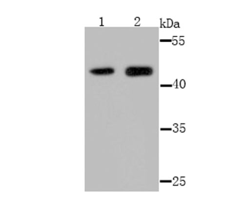
Fig1: Western blot analysis of Osteoprotegerin on 293 (1) and K562 (2) cell lysate using anti-Osteoprotegerin antibody at 1/500 dilution.Application
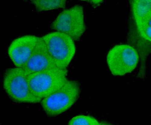
Fig2: ICC staining Osteoprotegerin in MCF-7 cells (green). The nuclear counter stain is DAPI (blue). Cells were fixed in paraformaldehyde, permeabilised with 0.25% Triton X100/PBS.Application
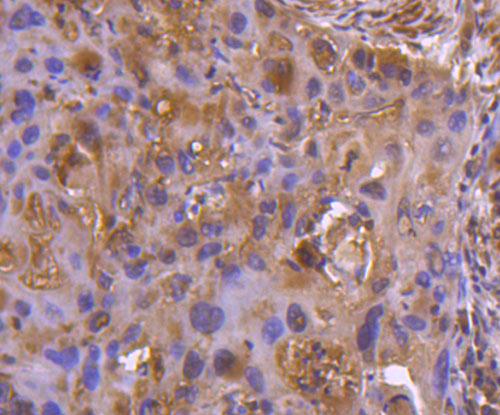
Fig3: Immunohistochemical analysis of paraffin-embedded human lung cancer tissue using anti-Osteoprotegerin antibody. Counter stained with hematoxylin.Application
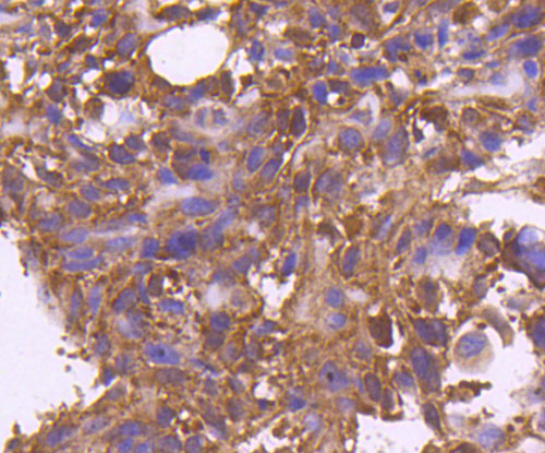
Fig4: Immunohistochemical analysis of paraffin-embedded human breast cancer tissue using anti-Osteoprotegerin antibody. Counter stained with hematoxylin.Application
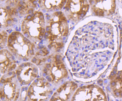
Fig5: Immunohistochemical analysis of paraffin-embedded human kidney tissue using anti-Osteoprotegerin antibody. Counter stained with hematoxylin.Application
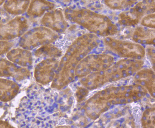
Fig6: Immunohistochemical analysis of paraffin-embedded mouse kidney tissue using anti-Osteoprotegerin antibody. Counter stained with hematoxylin.Application
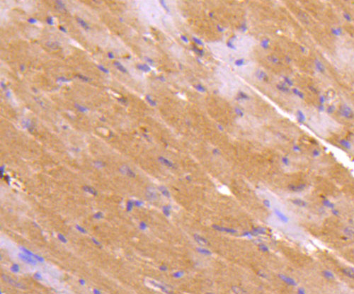
Fig7: Immunohistochemical analysis of paraffin-embedded mouse heart tissue using anti-Osteoprotegerin antibody. Counter stained with hematoxylin.| Product Name | Osteoprotegerin |
|---|---|
| Antibody Type | Primary Antibodies |
| Antigen Alias | MGC29565 antibody OCIF antibody OPG antibody Osteoclastogenesis inhibitory factor antibody Osteoprotegerin antibody PDB5 antibody TNF receptor superfamily member 11b antibody TNFRSF 11B antibody TNFRSF11B antibody TR 1 antibody TR1 antibody TR11B_HUMAN antibody Tumor necrosis factor receptor superfamily member 11B antibody |
| Product description | Bone morphogenesis and remodeling involve the formation of bone from osteoblasts and the resorption of bone by osteoclasts. The cytokine osteoprotegerin (OPG), also designated osteoclastogenesis inhibitory factor (OCIF), is known to inhibit osteoclast formation. A secreted glycoprotein, OPG is a member of the TNF receptor family that increases bone density and volume. OPG is thought to inhibit osteoclastogenesis by disrupting the cell-to-cell signaling between osteoblastic stromal cells and osteoclast progenitors. OPG is known to bind to TRAIL, a death domain-containing protein, and to inhibit TRAIL apoptosis in Jurkat cells. OPG also binds to osteoclast differentiation factor (ODF), also known as TRANCE/RANKL, a membrane-bound protein belonging to the TNF ligand family. Both TNFα and TNFβ upregulate OPG expression, while the bone resorbing agent prostaglandin E2 downregulates OPG. |
| Immunogen | Recombinant protein. |
| Clonality | Polyclonal |
|---|---|
| Isotype | IgG |
| Host Species | Rabbit |
| Tested Applications | WBICCIHC |
| WB:1:500~1:2000 ICC:1:200~1:500 IHC:1:50~1:200 Notes:Optimal dilutions/concentrations should be determined by the researcher. |
|
| Species Reactivity | HumanMouseRat |
| Concentration | 1mg/ml |
| Alternative Names | MGC29565 antibody
OCIF antibody OPG antibody Osteoclastogenesis inhibitory factor antibody Osteoprotegerin antibody PDB5 antibody TNF receptor superfamily member 11b antibody TNFRSF 11B antibody TNFRSF11B antibody TR 1 antibody TR1 antibody TR11B_HUMAN antibody Tumor necrosis factor receptor superfamily member 11B antibody |
|---|---|
| Molecular Weight(MW) | 46 kDa |
| Cellular Localization | Secreted. |
Application
Fig1: Western blot analysis of Osteoprotegerin on 293 (1) and K562 (2) cell lysate using anti-Osteoprotegerin antibody at 1/500 dilution.
Application
Fig2: ICC staining Osteoprotegerin in MCF-7 cells (green). The nuclear counter stain is DAPI (blue). Cells were fixed in paraformaldehyde, permeabilised with 0.25% Triton X100/PBS.
Application
Fig3: Immunohistochemical analysis of paraffin-embedded human lung cancer tissue using anti-Osteoprotegerin antibody. Counter stained with hematoxylin.
Application
Fig4: Immunohistochemical analysis of paraffin-embedded human breast cancer tissue using anti-Osteoprotegerin antibody. Counter stained with hematoxylin.
Application
Fig5: Immunohistochemical analysis of paraffin-embedded human kidney tissue using anti-Osteoprotegerin antibody. Counter stained with hematoxylin.
Application
Fig6: Immunohistochemical analysis of paraffin-embedded mouse kidney tissue using anti-Osteoprotegerin antibody. Counter stained with hematoxylin.
Application
Fig7: Immunohistochemical analysis of paraffin-embedded mouse heart tissue using anti-Osteoprotegerin antibody. Counter stained with hematoxylin.| Positive Control | 293, K562, MCF-7, human lung cancer tissue, human breast cancer tissue, human kidney tissue, mouse kidney tissue, mouse brain tissue. |
|---|---|
| Application Notes | WB:1:500~1:2000 ICC:1:200~1:500 IHC:1:50~1:200 Notes:Optimal dilutions/concentrations should be determined by the researcher. |
| Form | Liquid |
|---|---|
| Storage Instructions | Store at +4℃ after thawing. Aliquot store at -20℃ or -80℃. Avoid repeated freeze / thaw cycles. |
| Storage Buffer | 1*TBS (pH7.4), 0.5%BSA, 50%Glycerol. Preservative: 0.05% Sodium Azide. |


