Application
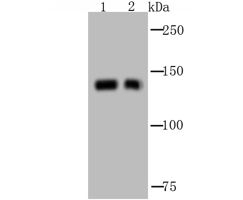
Fig1: Western blot analysis of GM130 on different cell lysate using anti-GM130 antibody at 1/1,000 dilution. Positive control: Lane1: MCF-7 Lane2: HelaApplication
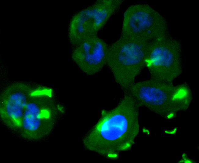
Fig2: ICC staining GM130 in LO2 cells (green). The nuclear counter stain is DAPI (blue). Cells were fixed in paraformaldehyde, permeabilised with 0.25% Triton X100/PBS.Application
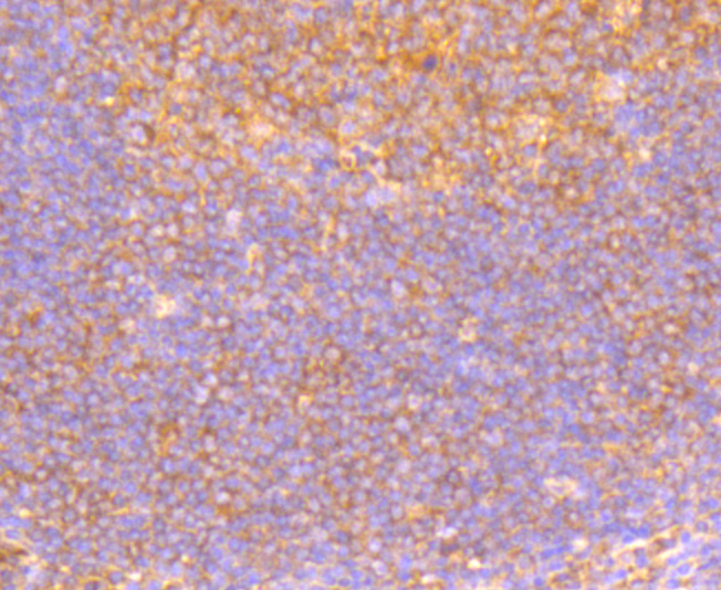
Fig3: Immunohistochemical analysis of paraffin-embedded human tonsil tissue using anti-GM130 antibody. Counter stained with hematoxylin.Application
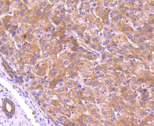
Fig4: Immunohistochemical analysis of paraffin-embedded human liver tissue using anti-GM130 antibody. Counter stained with hematoxylin.Application
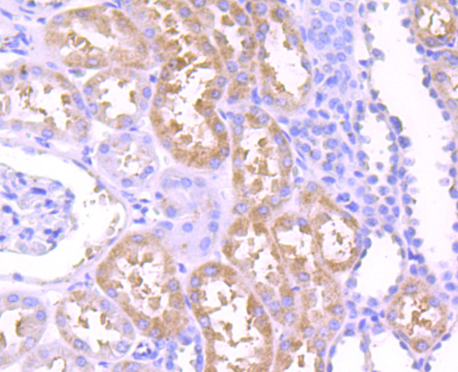
Fig5: Immunohistochemical analysis of paraffin-embedded human kidney tissue using anti-GM130 antibody. Counter stained with hematoxylin.Application
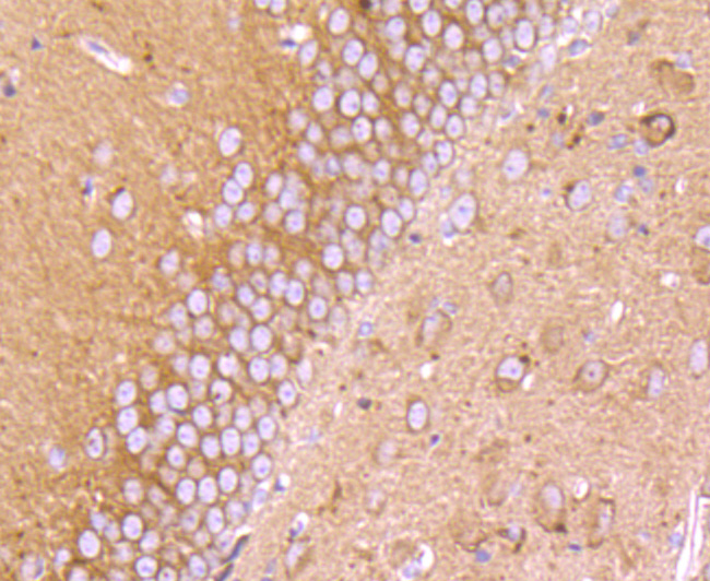
Fig6: Immunohistochemical analysis of paraffin-embedded mouse brain tissue using anti-GM130 antibody. Counter stained with hematoxylin.Application
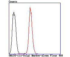
Fig7: Flow cytometric analysis of Hela cells with GM130 antibody at 1/100 dilution (red) compared with an unlabelled control (cells without incubation with primary antibody; black).| Product Name | GM130 (cis-Golgi Marker) |
|---|---|
| Antibody Type | Primary Antibodies |
| Antigen Alias | 30 kDa cis Golgi matrix protein antibody 130 kDa cis-Golgi matrix protein antibody Cis golgi matrix protein GM130 antibody GM130 antibody Gm130 autoantigen antibody GOGA2_HUMAN antibody GOLGA 2 antibody Golga2 antibody Golgi autoantigen antibody Golgi autoantigen golgin subfamily a 2 antibody Golgi matrix protein GM130 antibody Golgin 95 antibody golgin A2 antibody Golgin subfamily a 2 antibody Golgin subfamily A member 2 antibody Golgin-95 antibody MGC20672 antibody SY11 protein antibody |
| Product description | Peripheral membrane component of the cis-Golgi stack that acts as a membrane skeleton that maintains the structure of the Golgi apparatus, and as a vesicle thether that facilitates vesicle fusion to the Golgi membrane. Together with p115/USO1 and STX5, involved in vesicle tethering and fusion at the cis-Golgi membrane to maintain the stacked and inter-connected structure of the Golgi apparatus. Plays a central role in mitotic Golgi disassembly. Also plays a key role in spindle pole assembly and centrosome organization. Promotes the mitotic spindle pole assembly by activating the spindle assembly factor TPX2 to nucleate microtubules around the Golgi and capture them to couple mitotic membranes to the spindle. TPX2 then activates AURKA kinase and stimulates local microtubule nucleation. Upon filament assembly, nascent microtubules are further captured by GOLGA2, thus linking Golgi membranes to the spindle. Regulates the meiotic spindle pole assembly, probably via the same mechanism. Also regulates the centrosome organization. Also required for the Golgi ribbon formation and glycosylation of membrane and secretory proteins. |
| Immunogen | Recombinant protein |
| Clonality | Polyclonal |
|---|---|
| Isotype | IgG |
| Host Species | Rabbit |
| Tested Applications | WBICCIHCFC |
| WB:1:500-1:2,000 ICC:1:50-1:200 IHC:1:50-1:200 FC:1:50-1:200 |
|
| Species Reactivity | HumanMouseRat |
| Concentration | 1mg/ml |
| Alternative Names | 30 kDa cis Golgi matrix protein antibody 130 kDa cis-Golgi matrix protein antibody Cis golgi matrix protein GM130 antibody GM130 antibody Gm130 autoantigen antibody GOGA2_HUMAN antibody GOLGA 2 antibody Golga2 antibody Golgi autoantigen antibody Golgi autoantigen golgin subfamily a 2 antibody Golgi matrix protein GM130 antibody Golgin 95 antibody golgin A2 antibody Golgin subfamily a 2 antibody Golgin subfamily A member 2 antibody Golgin-95 antibody MGC20672 antibody SY11 protein antibody |
|---|---|
| Molecular Weight(MW) | 130 kDa |
| Cellular Localization | Cytoplasm, Cytoskeleton, Membrane, Microtubule. |
Application
Fig1: Western blot analysis of GM130 on different cell lysate using anti-GM130 antibody at 1/1,000 dilution. Positive control: Lane1: MCF-7 Lane2: Hela
Application
Fig2: ICC staining GM130 in LO2 cells (green). The nuclear counter stain is DAPI (blue). Cells were fixed in paraformaldehyde, permeabilised with 0.25% Triton X100/PBS.
Application
Fig3: Immunohistochemical analysis of paraffin-embedded human tonsil tissue using anti-GM130 antibody. Counter stained with hematoxylin.
Application
Fig4: Immunohistochemical analysis of paraffin-embedded human liver tissue using anti-GM130 antibody. Counter stained with hematoxylin.
Application
Fig5: Immunohistochemical analysis of paraffin-embedded human kidney tissue using anti-GM130 antibody. Counter stained with hematoxylin.
Application
Fig6: Immunohistochemical analysis of paraffin-embedded mouse brain tissue using anti-GM130 antibody. Counter stained with hematoxylin.
Application
Fig7: Flow cytometric analysis of Hela cells with GM130 antibody at 1/100 dilution (red) compared with an unlabelled control (cells without incubation with primary antibody; black).| Positive Control | Hela, MCF-7, LO2, human tonsil tissue, human liver tissue, human kidney tissue, mouse brain tissue. |
|---|---|
| Application Notes | WB:1:500-1:2,000 ICC:1:50-1:200 IHC:1:50-1:200 FC:1:50-1:200 |
| Form | Liquid |
|---|---|
| Storage Instructions | Store at +4℃ after thawing. Aliquot store at -20℃ or -80℃. Avoid repeated freeze / thaw cycles. |
| Storage Buffer | 1*TBS (pH7.4), 0.5%BSA, 50%Glycerol. Preservative: 0.05% Sodium Azide. |


