Application
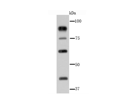
Fig1: Western blot analysis of Glucocorticoid Receptor alpha on Hela cell lysate using anti-Glucocorticoid Receptor alpha antibody at 1/1,000 dilution.Application
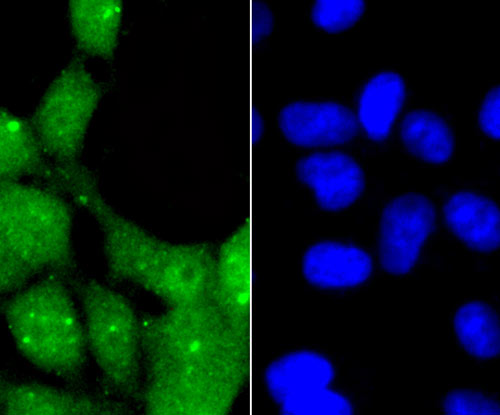
Fig2: ICC staining Glucocorticoid Receptor alpha in 293 cells (green). The nuclear counter stain is DAPI (blue). Cells were fixed in paraformaldehyde, permeabilised with 0.25% Triton X100/PBS.Application
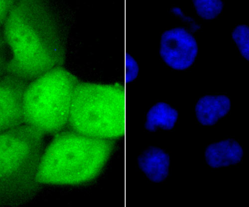
Fig3: ICC staining Glucocorticoid Receptor alpha in A431 cells (green). The nuclear counter stain is DAPI (blue). Cells were fixed in paraformaldehyde, permeabilised with 0.25% Triton X100/PBS.| Product Name | Glucocorticoid Receptor alpha |
|---|---|
| Antibody Type | Primary Antibodies |
| Antigen Alias | GCCR antibody GCR antibody Glucocorticoid receptor alpha isoform antibody Glucocorticoid receptor antibody GR antibody GRL antibody NR3C1 antibody Nuclear receptor subfamily 3 group C member 1 antibody |
| Product description | Glucocorticoids are a family of steroids necessary for the regulation of energy metabolism and the immune and inflammatory responses. These compounds exert their effect through their interaction with the Glucocorticoid Receptor (GR) and that complex's subsequent association with DNA. All normal mammalian tissues examined to date have been shown to contain GR. The human GR exists in two forms, alpha and beta, which are thought to be the result of alternative splicing of a single gene. Sequence analysis indicates that alpha and beta forms of human GR are 777 and 742 amino acids long, respectively. They are identical up to residue 727, after which they diverge. After ligand binding, the 94 kDa GR alpha isoform translocates from the cytoplasm to the nucleus where it regulates gene expression. In contrast, the 90 kDa GR beta isoform does not appear to bind either glucocorticoid agonists or antagonists, and has been localized predominantly in the nucleus independent of hormone treatment in some human cell lines. Studies suggest that human GR alpha has a greater affinity for GR response elements (GREs) than GR beta only when in the ligand bound state. |
| Immunogen | Recombinant protein. |
| Clonality | Polyclonal |
|---|---|
| Isotype | IgG |
| Host Species | Rabbit |
| Tested Applications | WBICC |
| WB:1:500~1:2000 ICC:1:200~1:500 Notes:Optimal dilutions/concentrations should be determined by the researcher. |
|
| Species Reactivity | HumanMouseRat |
| Concentration | 1mg/ml |
| Alternative Names | GCCR antibody GCR antibody Glucocorticoid receptor alpha isoform antibody Glucocorticoid receptor antibody GR antibody GRL antibody NR3C1 antibody Nuclear receptor subfamily 3 group C member 1 antibody |
|---|---|
| Molecular Weight(MW) | 86/76/60 kDa |
| Cellular Localization | Nucleus. Cytoplasm. |
Application
Fig1: Western blot analysis of Glucocorticoid Receptor alpha on Hela cell lysate using anti-Glucocorticoid Receptor alpha antibody at 1/1,000 dilution.
Application
Fig2: ICC staining Glucocorticoid Receptor alpha in 293 cells (green). The nuclear counter stain is DAPI (blue). Cells were fixed in paraformaldehyde, permeabilised with 0.25% Triton X100/PBS.
Application
Fig3: ICC staining Glucocorticoid Receptor alpha in A431 cells (green). The nuclear counter stain is DAPI (blue). Cells were fixed in paraformaldehyde, permeabilised with 0.25% Triton X100/PBS.| Positive Control | Hela, 293, A431. |
|---|---|
| Application Notes | WB:1:500~1:2000 ICC:1:200~1:500 Notes:Optimal dilutions/concentrations should be determined by the researcher. |
| Form | Liquid |
|---|---|
| Storage Instructions | Store at +4℃ after thawing. Aliquot store at -20℃ or -80℃. Avoid repeated freeze / thaw cycles. |
| Storage Buffer | 1*TBS (pH7.4), 0.5%BSA, 50%Glycerol. Preservative: 0.05% Sodium Azide. |


