Application
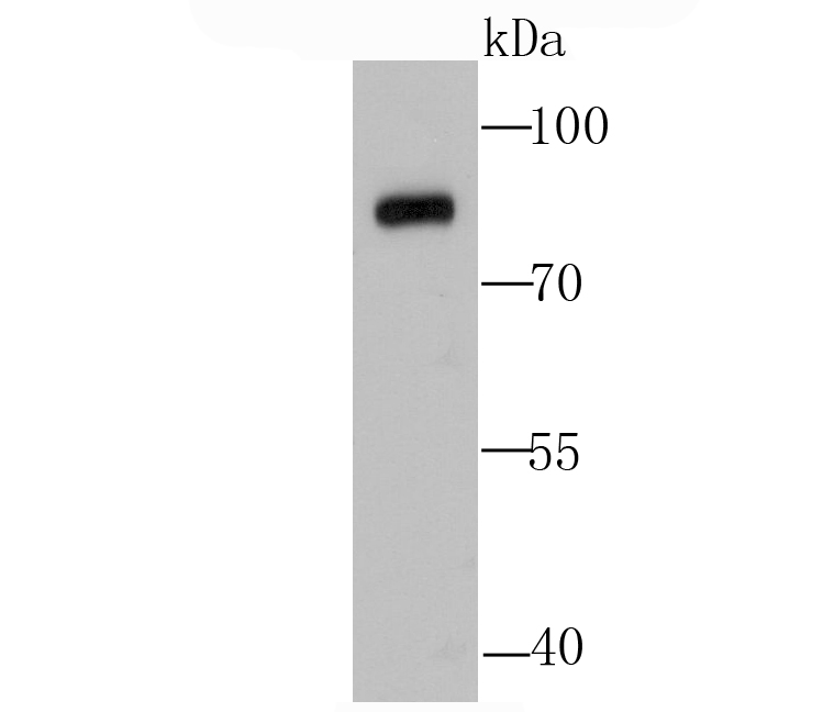
Fig1: Western blot analysis of Calpain 1 on HUVEC cell lysate using anti-Calpain 1 antibody at 1/500 dilution.Application
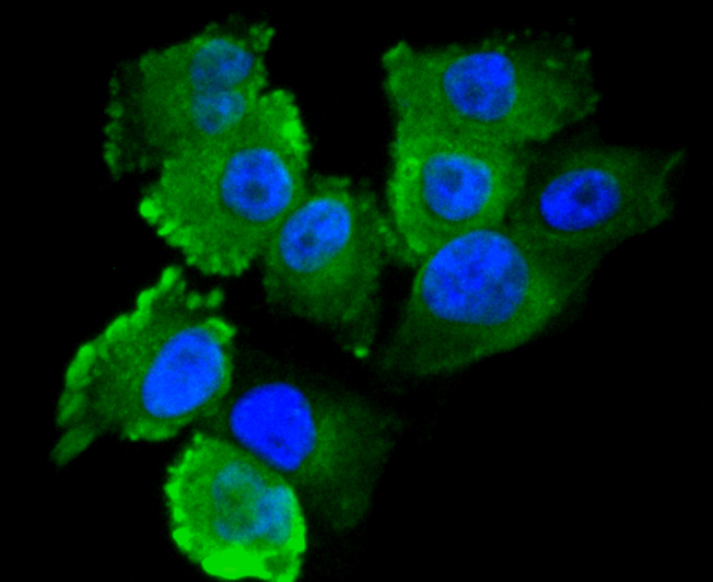
Fig2: ICC staining Calpain 1 in HUVEC cells (green). The nuclear counter stain is DAPI (blue). Cells were fixed in paraformaldehyde, permeabilised with 0.25% Triton X100/PBS.Application
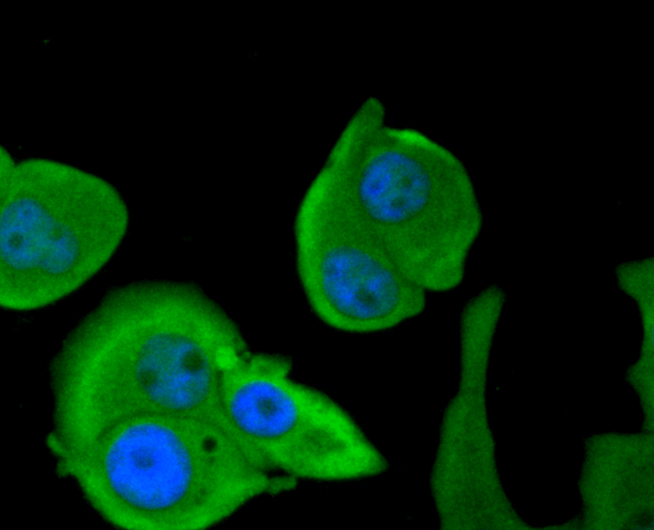
Fig3: ICC staining Calpain 1 in MCF-7 cells (green). The nuclear counter stain is DAPI (blue). Cells were fixed in paraformaldehyde, permeabilised with 0.25% Triton X100/PBS.Application
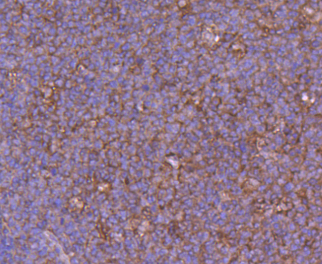
Fig4: Immunohistochemical analysis of paraffin-embedded human tonsil tissue using anti-Calpain 1 antibody. Counter stained with hematoxylin.Application
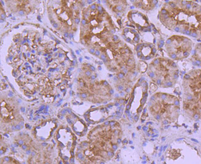
Fig5: Immunohistochemical analysis of paraffin-embedded human kidney tissue using anti-Calpain 1 antibody. Counter stained with hematoxylin.Application
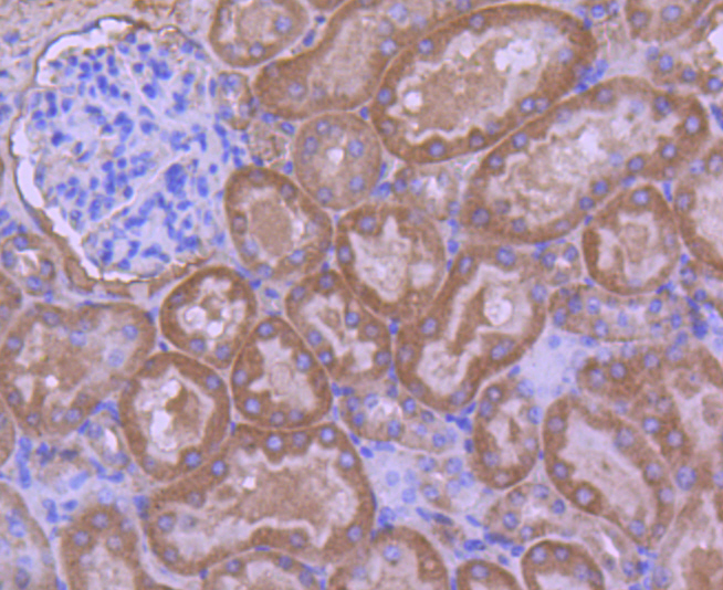
Fig6: Immunohistochemical analysis of paraffin-embedded 2 kidney tissue using anti-Calpain 1 antibody. Counter stained with hematoxylin.| Product Name | Calpain 1 |
|---|---|
| Antibody Type | Primary Antibodies |
| Antigen Alias | Ca2 activated neutral protease antibody Calcium activated neutral proteinase antibody Calcium activated neutral proteinase small subunit antibody Calcium dependent protease small subunit 1 antibody Calcium dependent protease small subunit antibody Calcium-activated neutral proteinase 1 antibody Calpain 1 antibody Calpain 1 large subunit antibody Calpain 1, (mu/I) large subunit antibody Calpain mu type antibody Calpain mu-type antibody Calpain regulatory subunit antibody Calpain small subunit 1 antibody Calpain, large polypeptide L1 antibody Calpain-1 catalytic subunit antibody Calpain-1 large subunit antibody CAN1_HUMAN antibody CANP 1 antibody CANP antibody CANP small subunit antibody CANP1 antibody CANPL 1 antibody CANPL1 antibody CAPN 1 antibody CAPN1 antibody Cell proliferation inducing protein 30 antibody Cell proliferation-inducing gene 30 protein antibody Micromolar Calpain antibody Micromolar-calpain antibody Mu Calpain antibody muCANP antibody muCL antibody |
| Product description | Calpain 1, also designated ?-calpain, is an intracellular calcium-dependent protease that cleaves cyto-skeletal and submembranous proteins. Calpains are nonlysosomal, calcium-activated intracellular cysteine proteases. Calpains mediate specific Ca2+-dependent processes including cell fusion, mitosis and meiosis. Calpains are heterodimers of a small regulatory subunit and one of three large catalytic subunits, designated Calpain 1, Calpain 2 and Calpain p94. Calpastatin regulates Calpain by inhibiting both the proteolytic activity of Calpain and its binding to membranes. Calpastatin exists in two types, tissue type and erythrocyte type, resulting from both alternative splicing and proteolytic processing. Calpain-1 co-localizes with human leukocyte antigen-DR (HLA-DR) on activated microglia in the aging brain. Calpain influences the process of spermatogenesis and the events preceding fertilization, such as the acrosome reaction. |
| Immunogen | Recombinant protein |
| Clonality | Polyclonal |
|---|---|
| Isotype | IgG |
| Host Species | Rabbit |
| Tested Applications | WBICCIHC |
| WB:1:500 ICC:1:50-1:200 IHC:1:50-1:200 |
|
| Species Reactivity | HumanMouseRat |
| Concentration | 1mg/ml |
| Alternative Names | Ca2 activated neutral protease antibody
Calcium activated neutral proteinase antibody
Calcium activated neutral proteinase small subunit antibody
Calcium dependent protease small subunit 1 antibody
Calcium dependent protease small subunit antibody
Calcium-activated neutral proteinase 1 antibody
Calpain 1 antibody
Calpain 1 large subunit antibody
Calpain 1 (mu/I) large subunit antibody Calpain mu type antibody Calpain mu-type antibody Calpain regulatory subunit antibody Calpain small subunit 1 antibody Calpain large polypeptide L1 antibody Calpain-1 catalytic subunit antibody Calpain-1 large subunit antibody CAN1_HUMAN antibody CANP 1 antibody CANP antibody CANP small subunit antibody CANP1 antibody CANPL 1 antibody CANPL1 antibody CAPN 1 antibody CAPN1 antibody Cell proliferation inducing protein 30 antibody Cell proliferation-inducing gene 30 protein antibody Micromolar Calpain antibody Micromolar-calpain antibody Mu Calpain antibody muCANP antibody muCL antibody |
|---|---|
| Molecular Weight(MW) | 82 kDa |
| Cellular Localization | Cytoplasm. Cell membrane. |
Application
Fig1: Western blot analysis of Calpain 1 on HUVEC cell lysate using anti-Calpain 1 antibody at 1/500 dilution.
Application
Fig2: ICC staining Calpain 1 in HUVEC cells (green). The nuclear counter stain is DAPI (blue). Cells were fixed in paraformaldehyde, permeabilised with 0.25% Triton X100/PBS.
Application
Fig3: ICC staining Calpain 1 in MCF-7 cells (green). The nuclear counter stain is DAPI (blue). Cells were fixed in paraformaldehyde, permeabilised with 0.25% Triton X100/PBS.
Application
Fig4: Immunohistochemical analysis of paraffin-embedded human tonsil tissue using anti-Calpain 1 antibody. Counter stained with hematoxylin.
Application
Fig5: Immunohistochemical analysis of paraffin-embedded human kidney tissue using anti-Calpain 1 antibody. Counter stained with hematoxylin.
Application
Fig6: Immunohistochemical analysis of paraffin-embedded 2 kidney tissue using anti-Calpain 1 antibody. Counter stained with hematoxylin.| Positive Control | HUVEC, MCF-7, human tonsil tissue, human kidney tissue, 2 kidney tissue. |
|---|---|
| Application Notes | WB:1:500 ICC:1:50-1:200 IHC:1:50-1:200 |
| Form | Liquid |
|---|---|
| Storage Instructions | Store at +4℃ after thawing. Aliquot store at -20℃ or -80℃. Avoid repeated freeze / thaw cycles. |
| Storage Buffer | 1*TBS (pH7.4), 1%BSA, 40%Glycerol. Preservative: 0.05% Sodium Azide. |


