Application
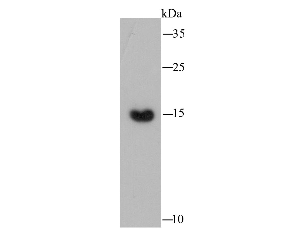
Fig1: Western blot analysis of COX IV on rat heart tissue lysate using anti-COX IV antibody at 1/2,000 dilution.Application
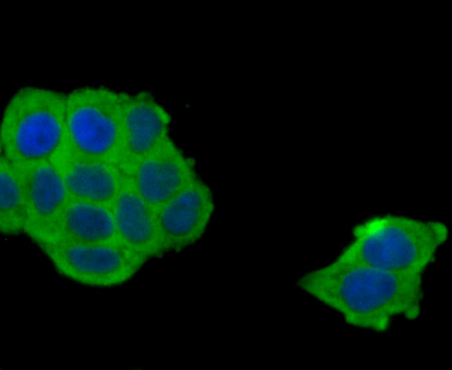
Fig2: ICC staining COX IV in Hela cells (green). The nuclear counter stain is DAPI (blue). Cells were fixed in paraformaldehyde, permeabilised with 0.25% Triton X100/PBS.Application
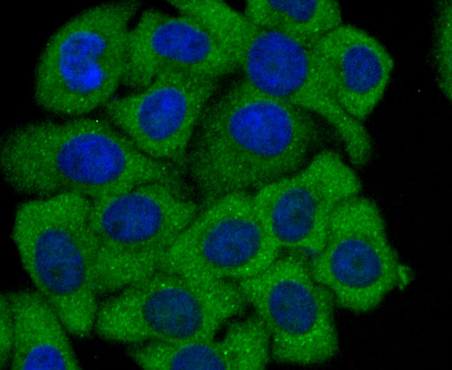
Fig3: ICC staining COX IV in HepG2 cells (green). The nuclear counter stain is DAPI (blue). Cells were fixed in paraformaldehyde, permeabilised with 0.25% Triton X100/PBS.Application
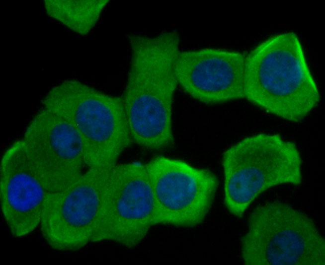
Fig4: ICC staining COX IV in MCF-7 cells (green). The nuclear counter stain is DAPI (blue). Cells were fixed in paraformaldehyde, permeabilised with 0.25% Triton X100/PBS.Application
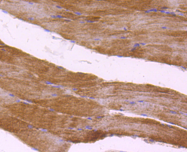
Fig5: Immunohistochemical analysis of paraffin-embedded rat skeletal muscle tissue using anti-COX IV antibody. Counter stained with hematoxylin.Application
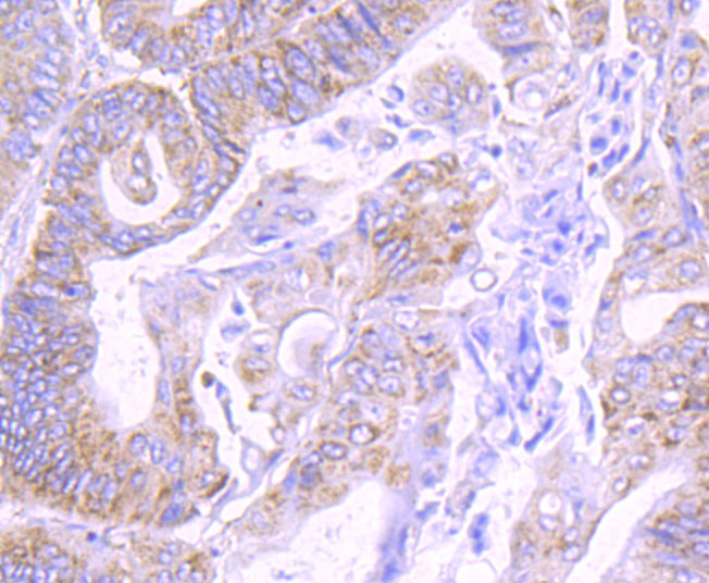
Fig6: Immunohistochemical analysis of paraffin-embedded human colon cancer tissue using anti-COX IV antibody. Counter stained with hematoxylin.Application
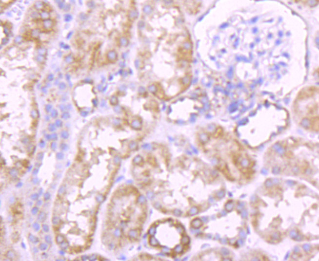
Fig7: Immunohistochemical analysis of paraffin-embedded human kidney tissue using anti-COX IV antibody. Counter stained with hematoxylin.Application
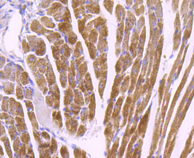
Fig8: Immunohistochemical analysis of paraffin-embedded mouse heart tissue using anti-COX IV antibody. Counter stained with hematoxylin.Application
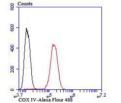
Fig9: Flow cytometric analysis of Hela cells with COX IV antibody at 1/100 dilution (red) compared with an unlabelled control (cells without incubation with primary antibody; black).| Product Name | COX IV |
|---|---|
| Antibody Type | Primary Antibodies |
| Antigen Alias | AL024441 antibody COX 4 antibody COX IV 1 antibody COX IV antibody COX IV-1 antibody Cox4 antibody COX41_HUMAN antibody Cox4a antibody COX4B antibody COX4I1 antibody COX4I2 antibody COX4L2 antibody COXIV antibody Cytochrome c oxidase polypeptide IV antibody Cytochrome c oxidase subunit 4 isoform 1 mitochondrial antibody Cytochrome c oxidase subunit 4 isoform 1, mitochondrial antibody Cytochrome C Oxidase subunit IV antibody Cytochrome c oxidase subunit IV isoform 1 antibody Cytochrome c oxidase subunit IV isoform 2 (lung) antibody Cytochrome c oxydase subunit 4 antibody dJ857M17.2 antibody MGC105470 antibody MGC72016 antibody |
| Product description | Cytochrome c oxidase (COX) functions as the terminal oxidase of the respiratory chain that uses cytochrome c as an electron donor to drive a proton gradient across the inner mitochondrial membrane. The mammalian COX apoenzyme is a heteromer consisting of three mitochondrial encoded catalytic subunits and several nuclear gene encoded structural subunits. COX contains two iron-coordination sites and two copper-coordination sites. Cytochrome c oxidase IV (COX4) is a nuclear-encoded subunit of COX that may play a role in regulating COX activity. COX4 is expressed ubiquitously in adult human tissue with the strongest levels of expression in the pancreas and moderate expression levels in heart, skeletal muscle and placenta. |
| Immunogen | Peptide |
| Clonality | Polyclonal |
|---|---|
| Isotype | IgG |
| Host Species | Rabbit |
| Tested Applications | WBICCIHCFC |
| WB:1:1,000-1:2,000 ICC:1:100-1:500 IHC:1:50-1:200 FC:1:50-1:100 |
|
| Species Reactivity | HumanMouseRat |
| Concentration | 1mg/ml |
| Alternative Names | AL024441 antibody
COX 4 antibody
COX IV 1 antibody
COX IV antibody
COX IV-1 antibody
Cox4 antibody
COX41_HUMAN antibody
Cox4a antibody
COX4B antibody
COX4I1 antibody
COX4I2 antibody
COX4L2 antibody
COXIV antibody
Cytochrome c oxidase polypeptide IV antibody
Cytochrome c oxidase subunit 4 isoform 1 mitochondrial antibody
Cytochrome c oxidase subunit 4 isoform 1 mitochondrial antibody Cytochrome C Oxidase subunit IV antibody Cytochrome c oxidase subunit IV isoform 1 antibody Cytochrome c oxidase subunit IV isoform 2 (lung) antibody Cytochrome c oxydase subunit 4 antibody dJ857M17.2 antibody MGC105470 antibody MGC72016 antibody |
|---|---|
| Molecular Weight(MW) | 16 kDa |
| Cellular Localization | Mitochondrion inner membrane. |
Application
Fig1: Western blot analysis of COX IV on rat heart tissue lysate using anti-COX IV antibody at 1/2,000 dilution.
Application
Fig2: ICC staining COX IV in Hela cells (green). The nuclear counter stain is DAPI (blue). Cells were fixed in paraformaldehyde, permeabilised with 0.25% Triton X100/PBS.
Application
Fig3: ICC staining COX IV in HepG2 cells (green). The nuclear counter stain is DAPI (blue). Cells were fixed in paraformaldehyde, permeabilised with 0.25% Triton X100/PBS.
Application
Fig4: ICC staining COX IV in MCF-7 cells (green). The nuclear counter stain is DAPI (blue). Cells were fixed in paraformaldehyde, permeabilised with 0.25% Triton X100/PBS.
Application
Fig5: Immunohistochemical analysis of paraffin-embedded rat skeletal muscle tissue using anti-COX IV antibody. Counter stained with hematoxylin.
Application
Fig6: Immunohistochemical analysis of paraffin-embedded human colon cancer tissue using anti-COX IV antibody. Counter stained with hematoxylin.
Application
Fig7: Immunohistochemical analysis of paraffin-embedded human kidney tissue using anti-COX IV antibody. Counter stained with hematoxylin.
Application
Fig8: Immunohistochemical analysis of paraffin-embedded mouse heart tissue using anti-COX IV antibody. Counter stained with hematoxylin.
Application
Fig9: Flow cytometric analysis of Hela cells with COX IV antibody at 1/100 dilution (red) compared with an unlabelled control (cells without incubation with primary antibody; black).| Positive Control | Rat heart tissue, Hela, MCF-7, HepG2, human colon cancer tissue, human kidney tissue, mouse heart tissue. |
|---|---|
| Application Notes | WB:1:1,000-1:2,000 ICC:1:100-1:500 IHC:1:50-1:200 FC:1:50-1:100 |
| Form | Liquid |
|---|---|
| Storage Instructions | Store at +4℃after thawing. Aliquot store at -20℃or -80℃. Avoid repeated freeze / thaw cycles. |
| Storage Buffer | 1*TBS (pH7.4), 0.5%BSA, 50%Glycerol. Preservative: 0.05% Sodium Azide. |


