Application
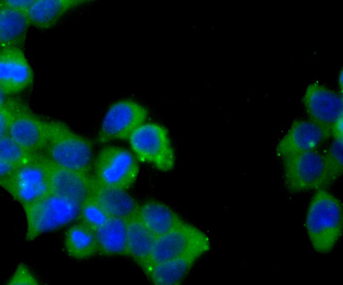
Fig1: ICC staining Jagged 1 in 293T cells (green). The nuclear counter stain is DAPI (blue). Cells were fixed in paraformaldehyde, permeabilised with 0.25% Triton X100/PBS.Application
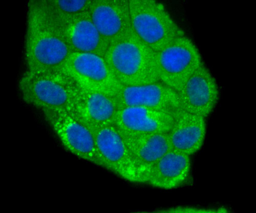
Fig2: ICC staining Jagged 1 in Hela cells (green). The nuclear counter stain is DAPI (blue). Cells were fixed in paraformaldehyde, permeabilised with 0.25% Triton X100/PBS.Application
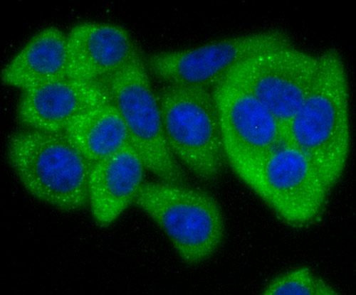
Fig3: ICC staining Jagged 1 in HepG2 cells (green). The nuclear counter stain is DAPI (blue). Cells were fixed in paraformaldehyde, permeabilised with 0.25% Triton X100/PBS.Application
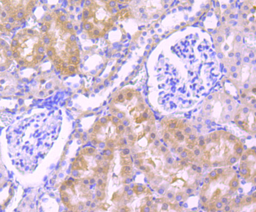
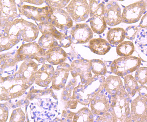
Fig4: Immunohistochemical analysis of paraffin-embedded rat kidney tissue using anti-Jagged 1 antibody. Counter stained with hematoxylin.Application
Fig5: Immunohistochemical analysis of paraffin-embedded mouse kidney tissue using anti-Jagged 1 antibody. Counter stained with hematoxylin.Application
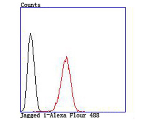
Fig6: Flow cytometric analysis of HepG2 cells with Jagged 1 antibody at 1/100 dilution (red) compared with an unlabelled control (cells without incubation with primary antibody; black).| Product Name | Jagged 1 |
|---|---|
| Antibody Type | Primary Antibodies |
| Antigen Alias | AGS antibody AHD antibody AWS antibody CD 339 antibody CD339 antibody CD339 antigen antibody Headturner antibody hJ1 antibody Htu antibody Jag 1 antibody Jag1 antibody JAG1_HUMAN antibody Jagged 1 antibody Jagged1 (Alagille syndrome) antibody Jagged1 antibody JAGL1 antibody MGC104644 antibody OTTHUMP00000030278 antibody Protein jagged-1 antibody Ser 1 antibody Ser1 antibody Serrate 1 antibody Slalom antibody |
| Product description | The LIN-12/Notch family of transmembrane receptors is believed to play a central role in development by regulating cell fate decisions. Ligands for Notch include Jagged1, Jagged2 and Delta. Jagged is a membrane protein and can activate Notch and prevent myoblast differentiation by inhibiting the expression of muscle regulatory and structural genes. It is involved in mammalian cardiovascular development and in cell-fate decisions during hematopoiesis. Jagged is expressed in adult and fetal tissues, and expression is upregulated in cervical squamous cell carcinoma. Familial Tetralogy of Fallot, the most common form of complex congenital heart disease, is caused by a mutation in the Jagged1 gene. |
| Immunogen | Peptide |
| Clonality | Polyclonal |
|---|---|
| Isotype | IgG |
| Host Species | Rabbit |
| Tested Applications | ICCIHCFC |
| ICC:1:200~1:500 IHC:1:50~1:200 FC:1:200~1:500 Notes:Optimal dilutions/concentrations should be determined by the researcher. |
|
| Species Reactivity | HumanMouseRat |
| Concentration | 1mg/ml |
| Alternative Names | AGS antibody AHD antibody AWS antibody CD 339 antibody CD339 antibody CD339 antigen antibody Headturner antibody hJ1 antibody Htu antibody Jag 1 antibody Jag1 antibody JAG1_HUMAN antibody Jagged 1 antibody Jagged1 (Alagille syndrome) antibody Jagged1 antibody JAGL1 antibody MGC104644 antibody OTTHUMP00000030278 antibody Protein jagged-1 antibody Ser 1 antibody Ser1 antibody Serrate 1 antibody Slalom antibody |
|---|---|
| Molecular Weight(MW) | 133 kDa |
| Cellular Localization | Membrane. |
Application
Fig1: ICC staining Jagged 1 in 293T cells (green). The nuclear counter stain is DAPI (blue). Cells were fixed in paraformaldehyde, permeabilised with 0.25% Triton X100/PBS.
Application
Fig2: ICC staining Jagged 1 in Hela cells (green). The nuclear counter stain is DAPI (blue). Cells were fixed in paraformaldehyde, permeabilised with 0.25% Triton X100/PBS.
Application
Fig3: ICC staining Jagged 1 in HepG2 cells (green). The nuclear counter stain is DAPI (blue). Cells were fixed in paraformaldehyde, permeabilised with 0.25% Triton X100/PBS.
Application
Fig4: Immunohistochemical analysis of paraffin-embedded rat kidney tissue using anti-Jagged 1 antibody. Counter stained with hematoxylin.
Application
Fig5: Immunohistochemical analysis of paraffin-embedded mouse kidney tissue using anti-Jagged 1 antibody. Counter stained with hematoxylin.
Application
Fig6: Flow cytometric analysis of HepG2 cells with Jagged 1 antibody at 1/100 dilution (red) compared with an unlabelled control (cells without incubation with primary antibody; black).| Positive Control | 293T, Hela, HepG2, rat kidney tissue, mouse kidney tissue. |
|---|---|
| Application Notes | ICC:1:200~1:500 IHC:1:50~1:200 FC:1:200~1:500 Notes:Optimal dilutions/concentrations should be determined by the researcher. |
| Form | Liquid |
|---|---|
| Storage Instructions | Store at +4℃ after thawing. Aliquot store at -20℃ or -80℃. Avoid repeated freeze / thaw cycles. |
| Storage Buffer | 1*TBS (pH7.4), 0.5%BSA, 50%Glycerol. Preservative: 0.05% Sodium Azide. |


