Application
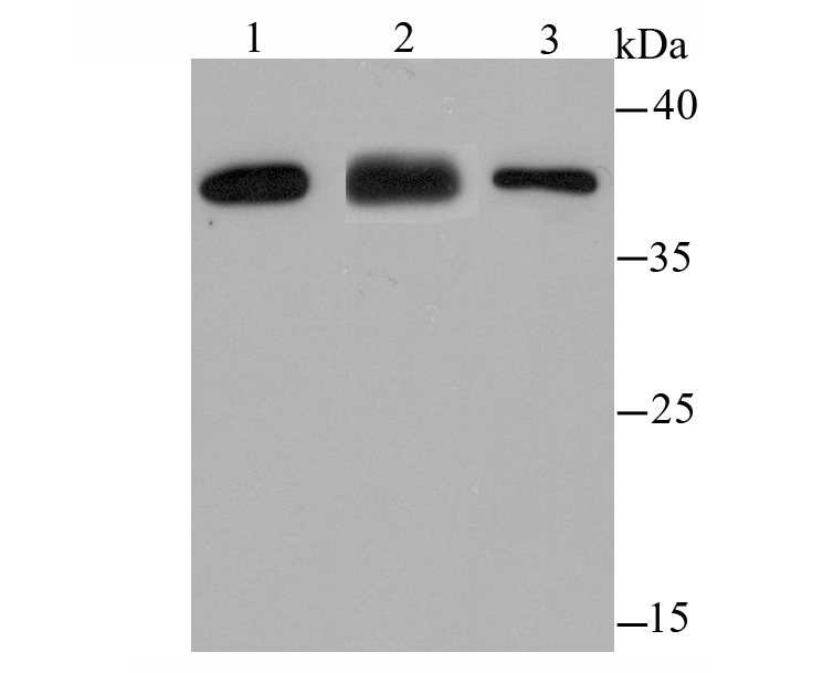
Fig1: Western blot analysis of IREB2 on different lysates using anti-IREB2 antibody at 1/500 dilution. Positive control: Lane1: K562 Lane2: Mouse kidney tissue Lane3: Rat skin tissueApplication
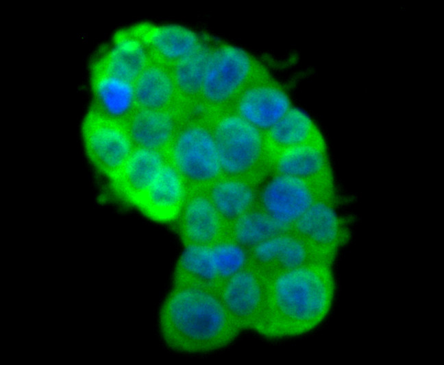
Fig2: ICC staining IREB2 in 293T cells (green). The nuclear counter stain is DAPI (blue). Cells were fixed in paraformaldehyde, permeabilised with 0.25% Triton X100/PBS.Application
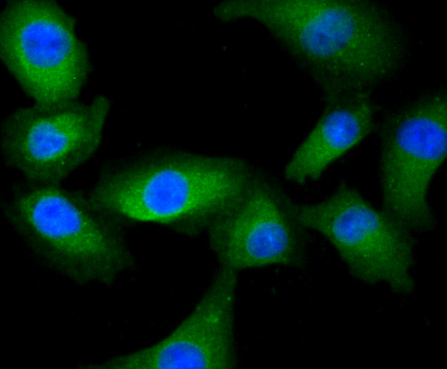
Fig3: ICC staining IREB2 in A549 cells (green). The nuclear counter stain is DAPI (blue). Cells were fixed in paraformaldehyde, permeabilised with 0.25% Triton X100/PBS.Application
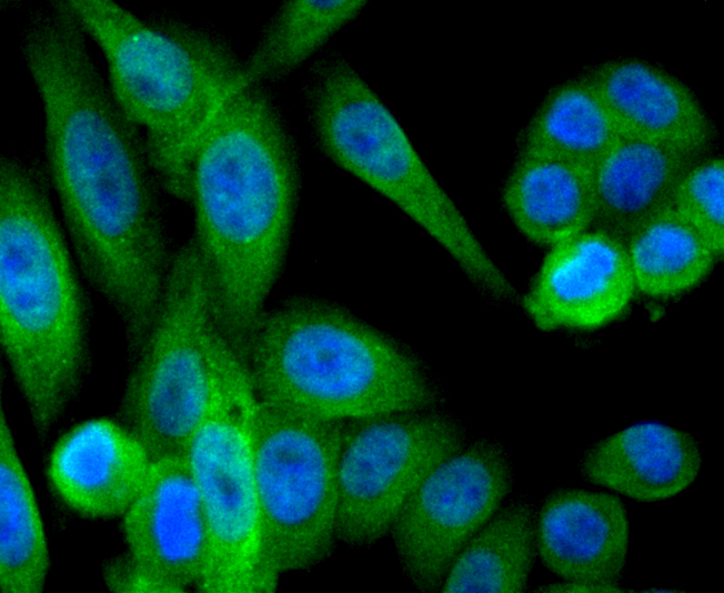
Fig4: ICC staining IREB2 in HepG2 cells (green). The nuclear counter stain is DAPI (blue). Cells were fixed in paraformaldehyde, permeabilised with 0.25% Triton X100/PBS.Application
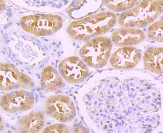
Fig5: Immunohistochemical analysis of paraffin-embedded human kidney tissue using anti-IREB2 antibody. Counter stained with hematoxylin.Application
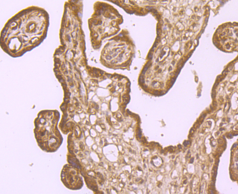
Fig6: Immunohistochemical analysis of paraffin-embedded human placenta tissue using anti-IREB2 antibody. Counter stained with hematoxylin.Application
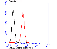
Fig7: Flow cytometric analysis of K562 cells with IREB2 antibody at 1/100 dilution (red) compared with an unlabelled control (cells without incubation with primary antibody; black).| Product Name | IREB2 |
|---|---|
| Antibody Type | Primary Antibodies |
| Antigen Alias | ACO3 antibody FLJ23381 antibody IRE BP2 antibody IRE-BP 2 antibody IREB 2 antibody IREB2 antibody IREB2_HUMAN antibody Iron regulatory protein 2 antibody iron responsive element binding protein 2 antibody Iron-responsive element-binding protein 2 antibody IRP 2 antibody IRP2 antibody IRP2AD antibody OTTHUMP00000185022 antibody |
| Product description | Iron metabolism is essential for sustaining mammalian homeostasis. Iron uptake and distribution is a highly regulated process in mammalian cells that is monitored by two iron sensing proteins: IRP-1 and -2 (iron regulatory protein-1 and -2), also known as iron responsive element-binding protein-1 and -2 (IRE-BP-1 and -2) or aconitase 1 and 2. IRP-1 and IRP-2 are important soluble regulatory factors that mediate iron uptake and storage in mammalian cells. They are capable of either repressing translation or enhancing mRNA stability by associating with stem-loop motifs known as iron-responsive elements (IREs). IRPs respond to stress mediators, iron concentration and signaling factors, including nitrogen monoxide, cytokines and hydrogen peroxide. |
| Immunogen | Recombinant protein |
| Clonality | Polyclonal |
|---|---|
| Isotype | IgG |
| Host Species | Rabbit |
| Tested Applications | WBICCIHCFC |
| WB:1:500-1:1000 ICC:1:50-1:200 IHC:1:50-1:200 FC:1:50-1:100 |
|
| Species Reactivity | HumanMouseRat |
| Concentration | 1mg/ml |
| Alternative Names | ACO3 antibody FLJ23381 antibody IRE BP2 antibody IRE-BP 2 antibody IREB 2 antibody IREB2 antibody IREB2_HUMAN antibody Iron regulatory protein 2 antibody iron responsive element binding protein 2 antibody Iron-responsive element-binding protein 2 antibody IRP 2 antibody IRP2 antibody IRP2AD antibody OTTHUMP00000185022 antibody |
|---|---|
| Molecular Weight(MW) | 105/37 kDa |
| Cellular Localization | Cytoplasm. |
Application
Fig1: Western blot analysis of IREB2 on different lysates using anti-IREB2 antibody at 1/500 dilution. Positive control: Lane1: K562 Lane2: Mouse kidney tissue Lane3: Rat skin tissue
Application
Fig2: ICC staining IREB2 in 293T cells (green). The nuclear counter stain is DAPI (blue). Cells were fixed in paraformaldehyde, permeabilised with 0.25% Triton X100/PBS.
Application
Fig3: ICC staining IREB2 in A549 cells (green). The nuclear counter stain is DAPI (blue). Cells were fixed in paraformaldehyde, permeabilised with 0.25% Triton X100/PBS.
Application
Fig4: ICC staining IREB2 in HepG2 cells (green). The nuclear counter stain is DAPI (blue). Cells were fixed in paraformaldehyde, permeabilised with 0.25% Triton X100/PBS.
Application
Fig5: Immunohistochemical analysis of paraffin-embedded human kidney tissue using anti-IREB2 antibody. Counter stained with hematoxylin.
Application
Fig6: Immunohistochemical analysis of paraffin-embedded human placenta tissue using anti-IREB2 antibody. Counter stained with hematoxylin.
Application
Fig7: Flow cytometric analysis of K562 cells with IREB2 antibody at 1/100 dilution (red) compared with an unlabelled control (cells without incubation with primary antibody; black).| Positive Control | K562, mouse kidney tissue lysate, rat skin tissue lysate, 293T, A549, HepG2, human kidney tissue, human placenta tissue. |
|---|---|
| Application Notes | WB:1:500-1:1000 ICC:1:50-1:200 IHC:1:50-1:200 FC:1:50-1:100 |
| Form | Liquid |
|---|---|
| Storage Instructions | Store at +4℃after thawing. Aliquot store at -20℃or -80℃. Avoid repeated freeze / thaw cycles. |
| Storage Buffer | 1*TBS (pH7.4), 0.5%BSA, 50%Glycerol. Preservative: 0.05% Sodium Azide. |


