Application
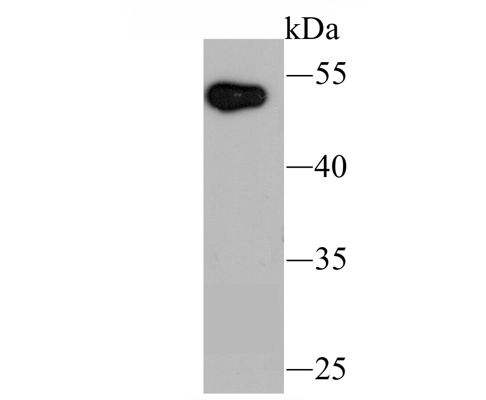
Fig1: Western blot analysis of UBE1C on human liver tissue lysate using anti-UBE1C antibody at 1/200 dilution.Application
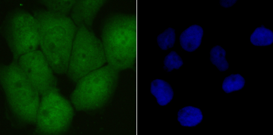
Fig2: ICC staining UBE1C in A431 cells (green). The nuclear counter stain is DAPI (blue). Cells were fixed in paraformaldehyde, permeabilised with 0.25% Triton X100/PBS.Application
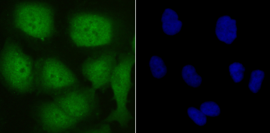
Fig3: ICC staining UBE1C in HUVEC cells (green). The nuclear counter stain is DAPI (blue). Cells were fixed in paraformaldehyde, permeabilised with 0.25% Triton X100/PBS.Application
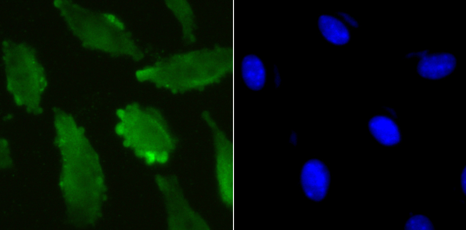
Fig4: ICC staining UBE1C in SH-SY5Y cells (green). The nuclear counter stain is DAPI (blue). Cells were fixed in paraformaldehyde, permeabilised with 0.25% Triton X100/PBS.Application
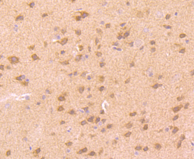
Fig5: Immunohistochemical analysis of paraffin-embedded rat brain tissue using anti-UBE1C antibody. Counter stained with hematoxylin.Application
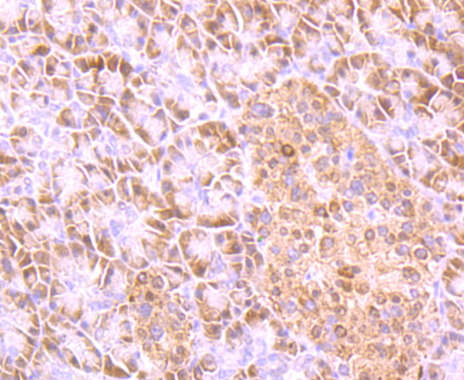
Fig6: Immunohistochemical analysis of paraffin-embedded human pancreas tissue using anti-UBE1C antibody. Counter stained with hematoxylin.Application
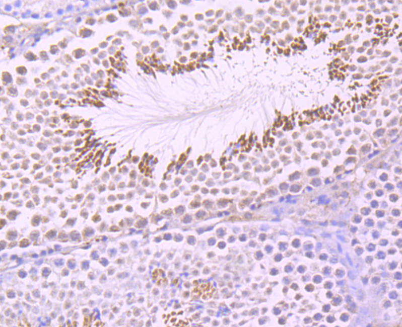
Fig7: Immunohistochemical analysis of paraffin-embedded mouse testis tissue using anti-UBE1C antibody. Counter stained with hematoxylin.Application
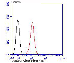
Fig8: Flow cytometric analysis of HUVEC cells with UBE1C antibody at 1/100 dilution (red) compared with an unlabelled control (cells without incubation with primary antibody; green).| Product Name | UBE1C |
|---|---|
| Antibody Type | Primary Antibodies |
| Antigen Alias | DKFZp566J164 antibody EC 6.3.2. antibody hUba3 antibody MGC22384 antibody NEDD8 activating enzyme E1 catalytic subunit antibody NEDD8 activating enzyme E1C antibody Nedd8 activating enzyme hUba3 antibody NEDD8-activating enzyme E1 catalytic subunit antibody NEDD8-activating enzyme E1C antibody uba3 antibody UBA3 ubiquitin activating enzyme E1 homolog antibody UBA3_HUMAN antibody UBE1C antibody Ubiquitin activating enzyme 3 antibody Ubiquitin activating enzyme E1C antibody Ubiquitin-activating enzyme 3 antibody Ubiquitin-activating enzyme E1C antibody Ubiquitin-like modifier-activating enzyme 3 antibody |
| Product description | Ubiquitin is an abundant, highly conserved protein found in all eukaryotic cells either free or covalently attached to cellular proteins. The primary function of ubiquitin in mammalian systems is to clear abnormal, foreign and improperly folded proteins by targeting them for proteosome degradation. UBA3 (ubiquitin-like modifier activating enzyme 3), also known as NEDD8-activating enzyme E1 catalytic subunit or UBE1C (ubiquitin-activating enzyme E1C), is a 463 amino acid protein belonging to the ubiquitin-activating E1 family and UBA3 subfamily. Ubiquitously expressed, UBA3 acts as an activator to NEDD8, a ubiquitin-like protein, thus regulating cell division, signaling and embryogenesis. UBA3 exists as two isoforms due to alternative splicing events. |
| Immunogen | Recombinant protein |
| Clonality | Polyclonal |
|---|---|
| Isotype | IgG |
| Host Species | Rabbit |
| Tested Applications | WBICCIHCFC |
| WB:1:500 ICC:1:50-1:200 IHC:1:50-1:200 FC:1:50-1:100 |
|
| Species Reactivity | HumanMouseRat |
| Concentration | 1mg/ml |
| Alternative Names | DKFZp566J164 antibody EC 6.3.2. antibody hUba3 antibody MGC22384 antibody NEDD8 activating enzyme E1 catalytic subunit antibody NEDD8 activating enzyme E1C antibody Nedd8 activating enzyme hUba3 antibody NEDD8-activating enzyme E1 catalytic subunit antibody NEDD8-activating enzyme E1C antibody uba3 antibody UBA3 ubiquitin activating enzyme E1 homolog antibody UBA3_HUMAN antibody UBE1C antibody Ubiquitin activating enzyme 3 antibody Ubiquitin activating enzyme E1C antibody Ubiquitin-activating enzyme 3 antibody Ubiquitin-activating enzyme E1C antibody Ubiquitin-like modifier-activating enzyme 3 antibody |
|---|---|
| Molecular Weight(MW) | 52 kDa |
| Cellular Localization | Nucleus. Cytoplasm. |
Application
Fig1: Western blot analysis of UBE1C on human liver tissue lysate using anti-UBE1C antibody at 1/200 dilution.
Application
Fig2: ICC staining UBE1C in A431 cells (green). The nuclear counter stain is DAPI (blue). Cells were fixed in paraformaldehyde, permeabilised with 0.25% Triton X100/PBS.
Application
Fig3: ICC staining UBE1C in HUVEC cells (green). The nuclear counter stain is DAPI (blue). Cells were fixed in paraformaldehyde, permeabilised with 0.25% Triton X100/PBS.
Application
Fig4: ICC staining UBE1C in SH-SY5Y cells (green). The nuclear counter stain is DAPI (blue). Cells were fixed in paraformaldehyde, permeabilised with 0.25% Triton X100/PBS.
Application
Fig5: Immunohistochemical analysis of paraffin-embedded rat brain tissue using anti-UBE1C antibody. Counter stained with hematoxylin.
Application
Fig6: Immunohistochemical analysis of paraffin-embedded human pancreas tissue using anti-UBE1C antibody. Counter stained with hematoxylin.
Application
Fig7: Immunohistochemical analysis of paraffin-embedded mouse testis tissue using anti-UBE1C antibody. Counter stained with hematoxylin.
Application
Fig8: Flow cytometric analysis of HUVEC cells with UBE1C antibody at 1/100 dilution (red) compared with an unlabelled control (cells without incubation with primary antibody; green).| Positive Control | Human liver tissue lysate, A431, HUVEC, SH-SY5Y, rat brain tissue, human pancreas tissue, mouse testis tissue. |
|---|---|
| Application Notes | WB:1:500 ICC:1:50-1:200 IHC:1:50-1:200 FC:1:50-1:100 |
| Form | Liquid |
|---|---|
| Storage Instructions | Store at +4℃after thawing. Aliquot store at -20℃or -80℃Avoid repeated freeze / thaw cycles. |
| Storage Buffer | 1*TBS (pH7.4), 0.5%BSA, 50%Glycerol. Preservative: 0.05% Sodium Azide. |


