Application
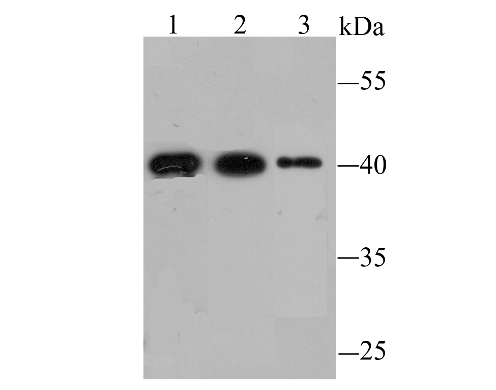
Fig1: Western blot analysis of FBXL2 on different lysates using anti-FBXL2 antibody at 1/1,000 dilution. Positive control: Lane1: Mouse liver tissue Lane2: Mouse heart tissue Lane3: SiHaApplication
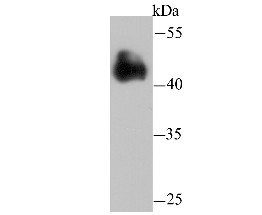
Fig2: Western blot analysis of FBXL2 on rat liver tissue lysate using anti-FBXL2 antibody at 1/10,000 dilution.Application
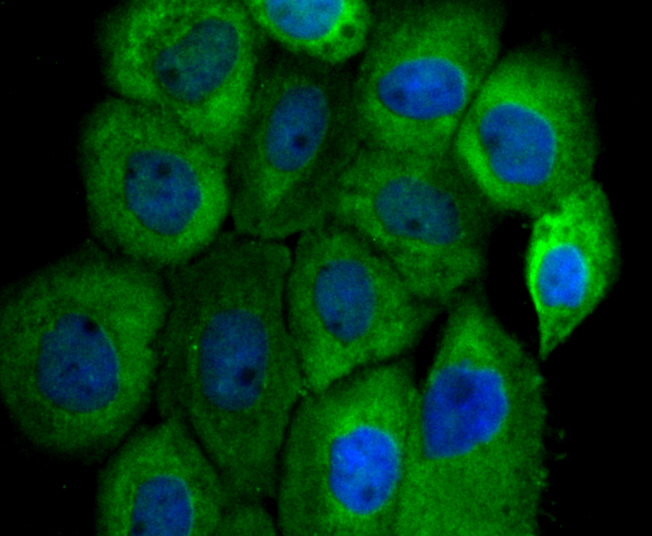
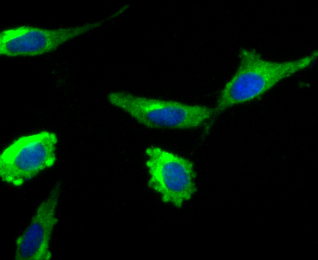
Fig3: ICC staining FBXL2 in A431 cells (green). The nuclear counter stain is DAPI (blue). Cells were fixed in paraformaldehyde, permeabilised with 0.25% Triton X100/PBS.Application
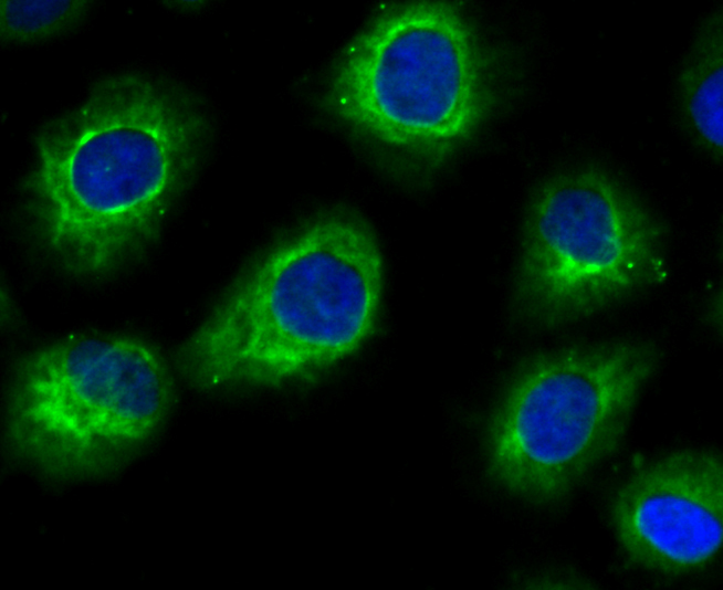
Fig4: ICC staining FBXL2 in HUVEC cells (green). The nuclear counter stain is DAPI (blue). Cells were fixed in paraformaldehyde, permeabilised with 0.25% Triton X100/PBS.Application
Fig5: ICC staining FBXL2 in SH-SY5Y cells (green). The nuclear counter stain is DAPI (blue). Cells were fixed in paraformaldehyde, permeabilised with 0.25% Triton X100/PBS.Application
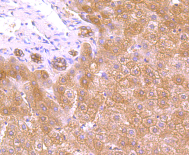
Fig6: Immunohistochemical analysis of paraffin-embedded human liver tissue using anti-FBXL2 antibody. Counter stained with hematoxylin.Application
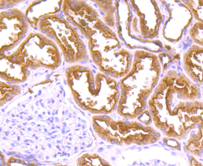
Fig7: Immunohistochemical analysis of paraffin-embedded human kidney tissue using anti-FBXL2 antibody. Counter stained with hematoxylin.Application
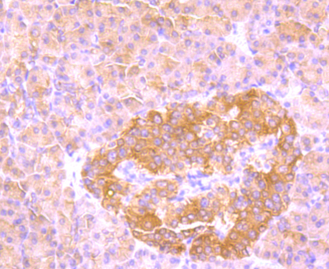
Fig8: Immunohistochemical analysis of paraffin-embedded human pancreas tissue using anti-FBXL2 antibody. Counter stained with hematoxylin.Application
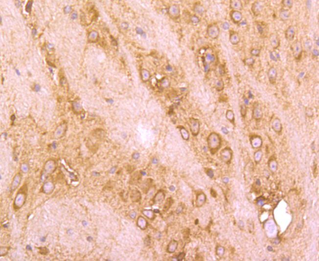
Fig9: Immunohistochemical analysis of paraffin-embedded mouse brain tissue using anti-FBXL2 antibody. Counter stained with hematoxylin.| Product Name | FBXL2 |
|---|---|
| Antibody Type | Primary Antibodies |
| Antigen Alias | DKFZP564P0622 antibody F box and leucine rich repeat protein 2 antibody F box protein containing leucine rich repeats antibody F box protein FBL2/FBL3 antibody F box/LRR repeat protein 2 antibody F-box and leucine-rich repeat protein 2 antibody F-box protein FBL2/FBL3 antibody F-box/LRR-repeat protein 2 antibody FBL 2 antibody FBL 3 antibody FBL2 antibody FBL3 antibody FBXL 2 antibody FBXL2 antibody FBXL2_HUMAN antibody |
| Product description | Calcium-activated substrate recognition component of a SCF (SKP1-cullin-F-box protein) E3 ubiquitin-protein ligase complex which mediates the ubiquitination and subsequent proteasomal degradation of target proteins. Unlike many F-box proteins, FBXL2 doesn't seem to target phosphodegron within its substrates but rather calmodulin-binding motifs. Targets PCYT1A for its monoubiquitination and degradation, this is antagonized by calmodulin (By similarity). Targets the cyclins CCND2 and CCND3 for polyubiquitination and degradation, leading to cell-cycle arrest in G(0), also antagonized by calmodulin. Binds to hepatitis C virus non-structural protein 5A (NS5A) in a reaction crucial for hepatitis C virus RNA replication. |
| Immunogen | Recombinant protein |
| Clonality | Polyclonal |
|---|---|
| Isotype | IgG |
| Host Species | Rabbit |
| Tested Applications | WBICCIHC |
| WB:1:1,000-1:10,000 ICC:1:50-1:200 IHC:1:50-1:200 |
|
| Species Reactivity | HumanMouseRat |
| Concentration | 1mg/ml |
| Alternative Names | DKFZP564P0622 antibody F box and leucine rich repeat protein 2 antibody F box protein containing leucine rich repeats antibody F box protein FBL2/FBL3 antibody F box/LRR repeat protein 2 antibody F-box and leucine-rich repeat protein 2 antibody F-box protein FBL2/FBL3 antibody F-box/LRR-repeat protein 2 antibody FBL 2 antibody FBL 3 antibody FBL2 antibody FBL3 antibody FBXL 2 antibody FBXL2 antibody FBXL2_HUMAN antibody |
|---|---|
| Molecular Weight(MW) | 49 kDa |
| Cellular Localization | Membrane. |
| SwissProt ID | Q9UKC9 |
|---|
Application
Fig1: Western blot analysis of FBXL2 on different lysates using anti-FBXL2 antibody at 1/1,000 dilution. Positive control: Lane1: Mouse liver tissue Lane2: Mouse heart tissue Lane3: SiHa
Application
Fig2: Western blot analysis of FBXL2 on rat liver tissue lysate using anti-FBXL2 antibody at 1/10,000 dilution.
Application
Fig3: ICC staining FBXL2 in A431 cells (green). The nuclear counter stain is DAPI (blue). Cells were fixed in paraformaldehyde, permeabilised with 0.25% Triton X100/PBS.
Application
Fig4: ICC staining FBXL2 in HUVEC cells (green). The nuclear counter stain is DAPI (blue). Cells were fixed in paraformaldehyde, permeabilised with 0.25% Triton X100/PBS.
Application
Fig5: ICC staining FBXL2 in SH-SY5Y cells (green). The nuclear counter stain is DAPI (blue). Cells were fixed in paraformaldehyde, permeabilised with 0.25% Triton X100/PBS.
Application
Fig6: Immunohistochemical analysis of paraffin-embedded human liver tissue using anti-FBXL2 antibody. Counter stained with hematoxylin.
Application
Fig7: Immunohistochemical analysis of paraffin-embedded human kidney tissue using anti-FBXL2 antibody. Counter stained with hematoxylin.
Application
Fig8: Immunohistochemical analysis of paraffin-embedded human pancreas tissue using anti-FBXL2 antibody. Counter stained with hematoxylin.
Application
Fig9: Immunohistochemical analysis of paraffin-embedded mouse brain tissue using anti-FBXL2 antibody. Counter stained with hematoxylin.| Positive Control | Mouse liver tissue lysate, mouse heart tissue lysate, SiHa, rat liver tissue lysate, A431, HUVEC, SH-SY5Y, human liver tissue, human kidney tissue, human pancreas tissue, mouse brain tissue. |
|---|---|
| Application Notes | WB:1:1,000-1:10,000 ICC:1:50-1:200 IHC:1:50-1:200 |
| Form | Liquid |
|---|---|
| Storage Instructions | Store at +4℃after thawing. Aliquot store at -20℃or -80℃. Avoid repeated freeze / thaw cycles. |
| Storage Buffer | 1*TBS (pH7.4), 0.5%BSA, 50%Glycerol. Preservative: 0.05% Sodium Azide. |


