Application
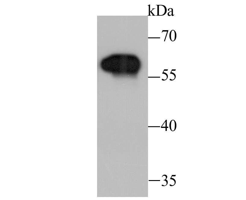
Fig1: Western blot analysis of Tyrosine Hydroxylase on PC-12 cell lysate using anti-Tyrosine Hydroxylase antibody at 1/1,000 dilution.Application
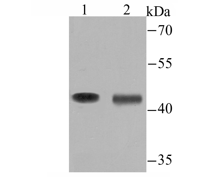
Fig2: Western blot analysis of Tyrosine Hydroxylase on mouse brain tissue (1) and MCF-7 cell (2) lysate using anti-Tyrosine Hydroxylase antibody at 1/500 dilution.Application
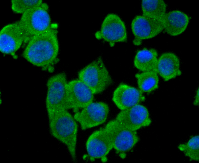
Fig3: ICC staining Tyrosine Hydroxylase in N2A cells (green). The nuclear counter stain is DAPI (blue). Cells were fixed in paraformaldehyde, permeabilised with 0.25% Triton X100/PBS.Application
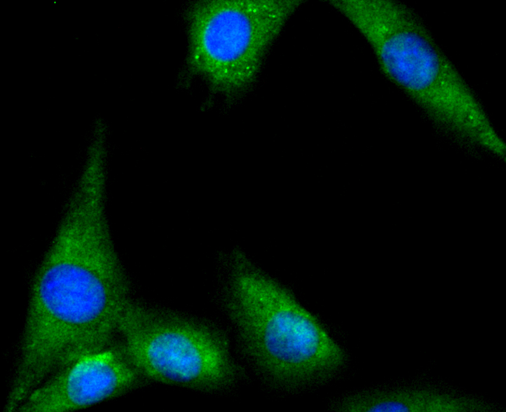
Fig4: ICC staining Tyrosine Hydroxylase in SHG-44 cells (green). The nuclear counter stain is DAPI (blue). Cells were fixed in paraformaldehyde, permeabilised with 0.25% Triton X100/PBS.Application
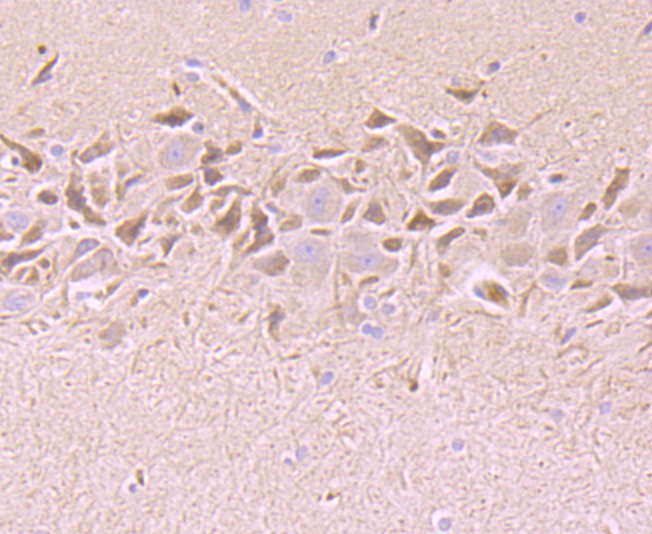
Fig5: Immunohistochemical analysis of paraffin-embedded rat brain tissue using anti-Tyrosine Hydroxylase antibody. Counter stained with hematoxylin.Application
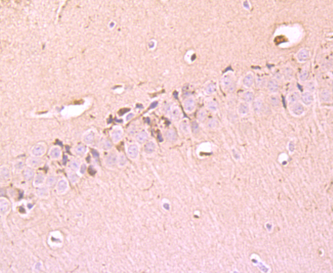
Fig6: Immunohistochemical analysis of paraffin-embedded mouse brain tissue using anti-Tyrosine Hydroxylase antibody. Counter stained with hematoxylin.Application
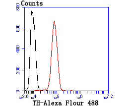
Fig7: Flow cytometric analysis of SH-SY5Y cells with Tyrosine Hydroxylase antibody at 1/100 dilution (red) compared with an unlabelled control (cells without incubation with primary antibody; black).| Product Name | Tyrosine Hydroxylase |
|---|---|
| Antibody Type | Primary Antibodies |
| Antigen Alias | Dystonia 14 antibody DYT14 antibody DYT5b antibody EC 1.14.16.2 antibody OTTHUMP00000011225 antibody OTTHUMP00000011226 antibody ple antibody Protein Pale antibody TH antibody The antibody TY3H_HUMAN antibody TYH antibody Tyrosine 3 hydroxylase antibody Tyrosine 3 monooxygenase antibody Tyrosine 3-hydroxylase antibody Tyrosine 3-monooxygenase antibody Tyrosine hydroxylase antibody |
| Product description | The enzyme tyrosine hydroxylase (TH), also designated tyrosine 3-monooxygenase (TY3H), catalyzes the conversion of tyrosine to L-dopa, which is the rate limiting step in the biosynthesis of catecholamines such as dopamine, adrenalin and noradrenalin. TH is thought to play a role in the pathogenesis of Parkinson's disease, which is associated with reduced dopamine levels. Two transcription factor binding sites in the proximal region of the TH gene, the TPA-responsive element (TRE) and the c-AMP responsive element (CRE), have been implicated in the complex regulation of the TH gene. TH is also known to be upregulated by the glia maturation factor (GMF), a Cdc 10/SWI6 motif-containing protein called V-1, and a variety of additional compounds. |
| Immunogen | Recombinant protein |
| Clonality | Polyclonal |
|---|---|
| Isotype | IgG |
| Host Species | Rabbit |
| Tested Applications | WBICCIHCFC |
| WB:1:1,000-1:2,000 ICC:1:50-1:200 IHC:1:50-1:200 FC:1:50-1:100 |
|
| Species Reactivity | HumanMouseRat |
| Concentration | 1mg/ml |
| Alternative Names | Dystonia 14 antibody DYT14 antibody DYT5b antibody EC 1.14.16.2 antibody OTTHUMP00000011225 antibody OTTHUMP00000011226 antibody ple antibody Protein Pale antibody TH antibody The antibody TY3H_HUMAN antibody TYH antibody Tyrosine 3 hydroxylase antibody Tyrosine 3 monooxygenase antibody Tyrosine 3-hydroxylase antibody Tyrosine 3-monooxygenase antibody Tyrosine hydroxylase antibody |
|---|---|
| Molecular Weight(MW) | 45/58 kDa |
| Cellular Localization | Cytoplasm. Nucleus. |
Application
Fig1: Western blot analysis of Tyrosine Hydroxylase on PC-12 cell lysate using anti-Tyrosine Hydroxylase antibody at 1/1,000 dilution.
Application
Fig2: Western blot analysis of Tyrosine Hydroxylase on mouse brain tissue (1) and MCF-7 cell (2) lysate using anti-Tyrosine Hydroxylase antibody at 1/500 dilution.
Application
Fig3: ICC staining Tyrosine Hydroxylase in N2A cells (green). The nuclear counter stain is DAPI (blue). Cells were fixed in paraformaldehyde, permeabilised with 0.25% Triton X100/PBS.
Application
Fig4: ICC staining Tyrosine Hydroxylase in SHG-44 cells (green). The nuclear counter stain is DAPI (blue). Cells were fixed in paraformaldehyde, permeabilised with 0.25% Triton X100/PBS.
Application
Fig5: Immunohistochemical analysis of paraffin-embedded rat brain tissue using anti-Tyrosine Hydroxylase antibody. Counter stained with hematoxylin.
Application
Fig6: Immunohistochemical analysis of paraffin-embedded mouse brain tissue using anti-Tyrosine Hydroxylase antibody. Counter stained with hematoxylin.
Application
Fig7: Flow cytometric analysis of SH-SY5Y cells with Tyrosine Hydroxylase antibody at 1/100 dilution (red) compared with an unlabelled control (cells without incubation with primary antibody; black).| Positive Control | PC-12, mouse brain tissue, MCF-7, N2A, SHG-44, rat brain tissue, SH-SY5Y. |
|---|---|
| Application Notes | WB:1:1,000-1:2,000 ICC:1:50-1:200 IHC:1:50-1:200 FC:1:50-1:100 |
| Form | Liquid |
|---|---|
| Storage Instructions | Store at +4℃after thawing. Aliquot store at -20℃or -80℃. Avoid repeated freeze / thaw cycles. |
| Storage Buffer | 1*TBS (pH7.4), 0.5%BSA, 50%Glycerol. Preservative: 0.05% Sodium Azide. |


