Application
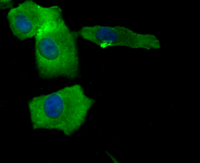
Fig1: ICC staining BAX in A549 cells (green). The nuclear counter stain is DAPI (blue). Cells were fixed in paraformaldehyde, permeabilised with 0.25% Triton X100/PBS.Application
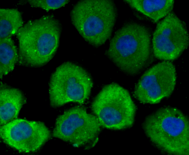
Fig2: ICC staining BAX in HepG2 cells (green). The nuclear counter stain is DAPI (blue). Cells were fixed in paraformaldehyde, permeabilised with 0.25% Triton X100/PBS.Application
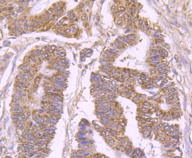
Fig3: Immunohistochemical analysis of paraffin-embedded human colon cancer tissue using anti-BAX antibody. Counter stained with hematoxylin.Application
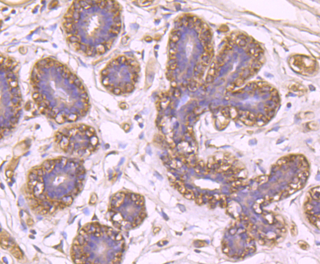
Fig4: Immunohistochemical analysis of paraffin-embedded human breast tissue using anti-BAX antibody. Counter stained with hematoxylin.Application
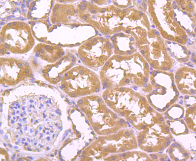
Fig5: Immunohistochemical analysis of paraffin-embedded human kidney tissue using anti-BAX antibody. Counter stained with hematoxylin.Application
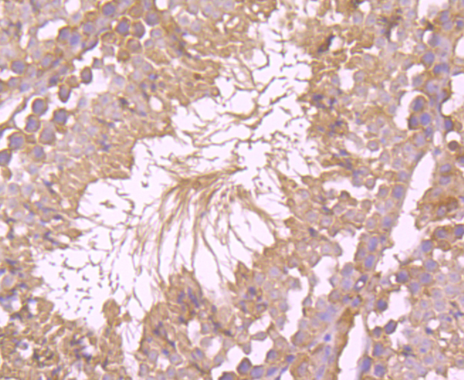
Fig6: Immunohistochemical analysis of paraffin-embedded mouse testis tissue using anti-BAX antibody. Counter stained with hematoxylin.| Product Name | BAX |
|---|---|
| Antibody Type | Primary Antibodies |
| Antigen Alias | Apoptosis regulator BAX antibody BAX antibody Bax-protein antibody BAX_HUMAN antibody BAXA antibody Baxdelta2G9 antibody Baxdelta2G9omega antibody Baxdelta2omega antibody Bcl-2-like protein 4 antibody BCL2 associated X protein antibody BCL2 associated X protein omega antibody BCL2 associated X protein transcript variant delta2 antibody Bcl2-L-4 antibody BCL2L4 antibody membrane isoform alpha antibody |
| Product description | Bax is a key component for cellular induced apoptosis through mitochondrial stress. It accelerates programmed cell death by binding to, and antagonizing the apoptosis repressor BCL2 or its adenovirus homolog E1B 19k protein. Under stress conditions, BAX undergoes a conformation change that causes translocation to the mitochondrion membrane, leading to the release of cytochrome c that then triggers apoptosis. It promotes activation of CASP3, and thereby apoptosis. |
| Immunogen | Peptide-KLH |
| Clonality | Polyclonal |
|---|---|
| Isotype | IgG |
| Host Species | Rabbit |
| Tested Applications | ICCIHC |
| ICC:1:100-1:200 IHC:1:50-1:200 |
|
| Species Reactivity | HumanMouse |
| Concentration | 1mg/ml |
| Alternative Names | Apoptosis regulator BAX antibody BAX antibody Bax-protein antibody BAX_HUMAN antibody BAXA antibody Baxdelta2G9 antibody Baxdelta2G9omega antibody Baxdelta2omega antibody Bcl-2-like protein 4 antibody BCL2 associated X protein antibody BCL2 associated X protein omega antibody BCL2 associated X protein transcript variant delta2 antibody Bcl2-L-4 antibody BCL2L4 antibody membrane isoform alpha antibody |
|---|---|
| Molecular Weight(MW) | 21 kDa |
| Cellular Localization | Mitochondrion membrane, cytoplasm. |
Application
Fig1: ICC staining BAX in A549 cells (green). The nuclear counter stain is DAPI (blue). Cells were fixed in paraformaldehyde, permeabilised with 0.25% Triton X100/PBS.
Application
Fig2: ICC staining BAX in HepG2 cells (green). The nuclear counter stain is DAPI (blue). Cells were fixed in paraformaldehyde, permeabilised with 0.25% Triton X100/PBS.
Application
Fig3: Immunohistochemical analysis of paraffin-embedded human colon cancer tissue using anti-BAX antibody. Counter stained with hematoxylin.
Application
Fig4: Immunohistochemical analysis of paraffin-embedded human breast tissue using anti-BAX antibody. Counter stained with hematoxylin.
Application
Fig5: Immunohistochemical analysis of paraffin-embedded human kidney tissue using anti-BAX antibody. Counter stained with hematoxylin.
Application
Fig6: Immunohistochemical analysis of paraffin-embedded mouse testis tissue using anti-BAX antibody. Counter stained with hematoxylin.| Positive Control | A549, HepG2, human breast tissue, human colon cancer tissue, human kidney tissue, mouse testis tissue. |
|---|---|
| Application Notes | ICC:1:100-1:200 IHC:1:50-1:200 |
| Form | Liquid |
|---|---|
| Storage Instructions | Store at +4℃after thawing. Aliquot store at -20℃or -80℃. Avoid repeated freeze / thaw cycles. |
| Storage Buffer | 1*TBS (pH7.4), 0.5%BSA, 50%Glycerol. Preservative: 0.05% Sodium Azide. |


