Application
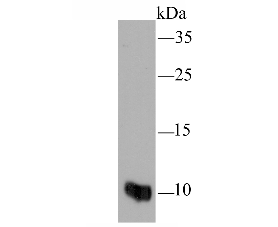
Fig1: Western blot analysis of MRP8/S100A8 on HL-60 cell lysate using anti-MRP8/S100A8 antibody at 1/100 dilution.Application
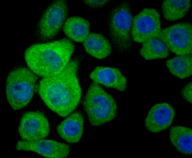
Fig2: ICC staining MRP8/S100A8 in AGS cells (green). The nuclear counter stain is DAPI (blue). Cells were fixed in paraformaldehyde, permeabilised with 0.25% Triton X100/PBS.Application
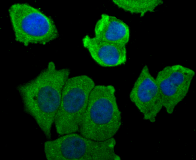
Fig3: ICC staining MRP8/S100A8 in MCF-7 cells (green). The nuclear counter stain is DAPI (blue). Cells were fixed in paraformaldehyde, permeabilised with 0.25% Triton X100/PBS.Application
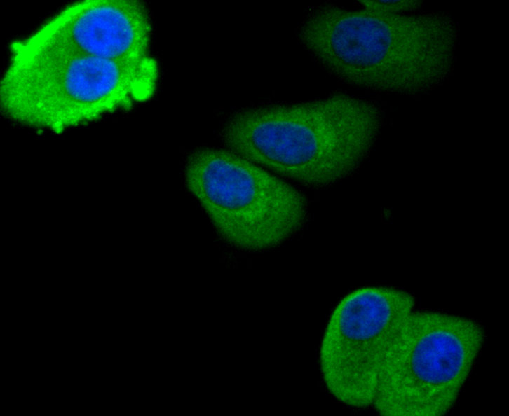
Fig4: ICC staining MRP8/S100A8 in SK-Br-3 cells (green). The nuclear counter stain is DAPI (blue). Cells were fixed in paraformaldehyde, permeabilised with 0.25% Triton X100/PBS.Application
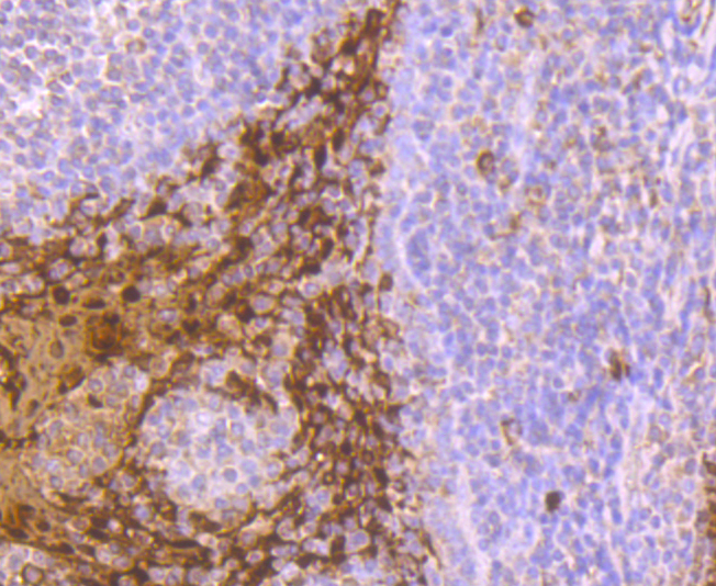
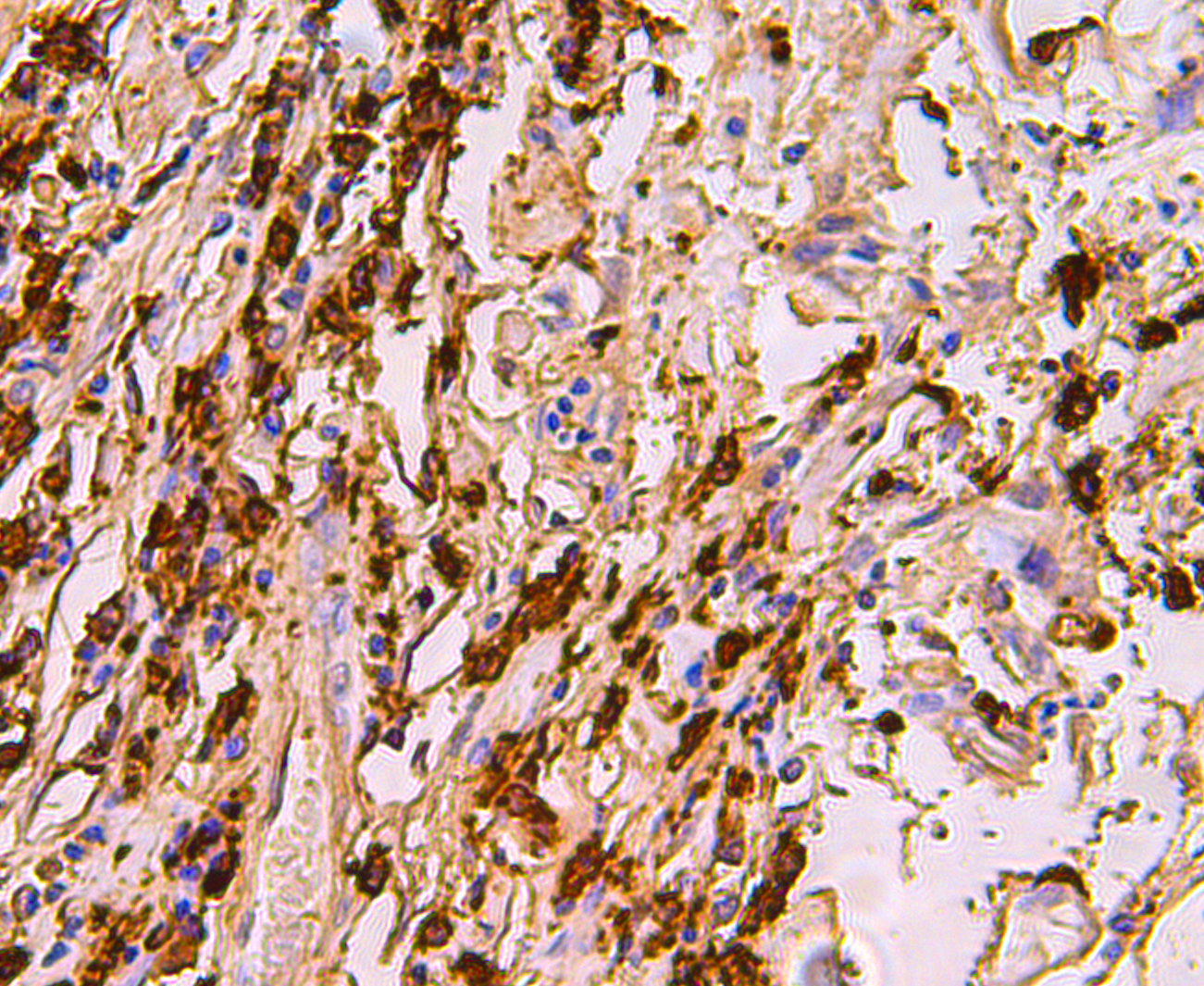
Fig5: Immunohistochemical analysis of paraffin-embedded human tonsil tissue using anti-MRP8/S100A8 antibody. Counter stained with hematoxylin.Application
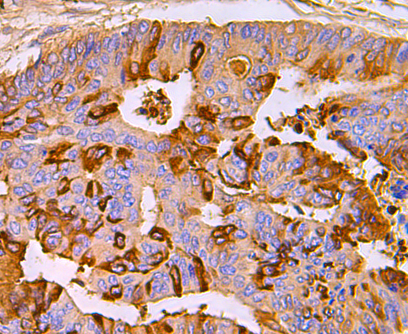
Fig6: Immunohistochemical analysis of paraffin-embedded human colon cancer tissue using anti-MRP8/S100A8 antibody. Counter stained with hematoxylin.Application
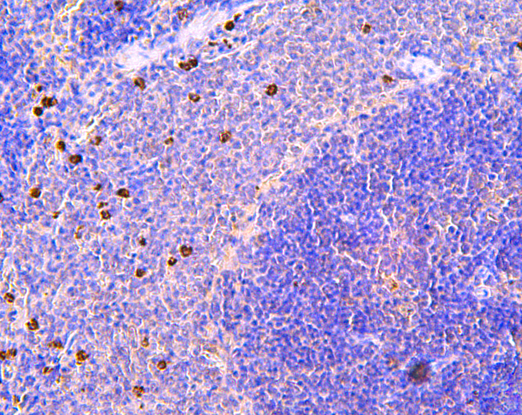
Fig7: Immunohistochemical analysis of paraffin-embedded rat spleen tissue using anti-MRP8/S100A8 antibody. Counter stained with hematoxylin.Application
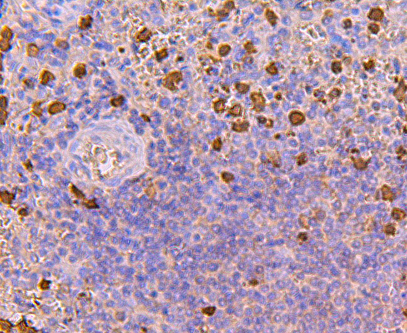
Fig8: Immunohistochemical analysis of paraffin-embedded human spleen tissue using anti-MRP8/S100A8 antibody. Counter stained with hematoxylin.Application
Fig9: Immunohistochemical analysis of paraffin-embedded human breast cancer tissue using anti-MRP8/S100A8 antibody. Counter stained with hematoxylin.Application
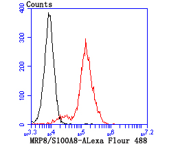
Fig10: Flow cytometric analysis of THP-1 cells with MRP8/S100A8 antibody at 1/100 dilution (red) compared with an unlabelled control (cells without incubation with primary antibody; black).| Product Name | MRP8/S100A8 |
|---|---|
| Antibody Type | Primary Antibodies |
| Antigen Alias | 60B8Ag antibody AI323541 antibody B8Ag antibody BEE11 antibody CAGA antibody Calgranulin-A antibody Calprotectin L1L subunit antibody Calprotectin, included antibody CFAG antibody CGLA antibody Chemotactic cytokine CP-10 antibody CP-10 antibody Cystic fibrosis antigen antibody L1Ag antibody Leukocyte L1 complex light chain antibody MA387 antibody MIF antibody Migration inhibitory factor-related protein 8 antibody MRP-8 antibody Myeloid-related protein 8 antibody Neutrophil cytosolic 7 kDa protein antibody NIF antibody p8 antibody Pro-inflammatory S100 cytokine antibody Protein S100-A8 antibody S100 calcium binding protein A8 (calgranulin A) antibody S100 calcium binding protein A8 antibody S100 calcium-binding protein A8 antibody S100A8 antibody S100A8/S100A9 complex, included antibody S10A8_HUMAN antibody Urinary stone protein band A antibody |
| Product description | The family of EF-hand type Ca2+-binding proteins includes Calbindin (previously designated vitamin D-dependent Ca2+-binding protein), S-100 α and β, Cal-granulins A (also designated MRP8), B (also designated MRP14) and C (S-100 like proteins) and the parvalbumin family members, including parvalbumin α and parvalbumin β (also designated oncomodulin). Calbindin, S-100 proteins and parvalbumin proteins are each expressed in neural tissues. In addition, S-100 α and β are present in a variety of other tissues and Calbindin is present in intestine and kidney. Parvalbumin α is also found in fast-contracting/ relaxing skeletal muscle fibers and parvalbumin β is found in many tumor tissues as well as in the organ of Corti. Calbindin, S-100 proteins and parvalbulmins have all been detected in Leydig cells and the testis. These proteins are thought to play a role in hormone production and spermatogenesis. Calgranulin is expressed in macrophages and epithelial cells. |
| Immunogen | Recombinant protein |
| Clonality | Polyclonal |
|---|---|
| Isotype | IgG |
| Host Species | Rabbit |
| Tested Applications | WBICCIHCFC |
| WB:1:500 ICC:1:50-1:200 IHC:1:50-1:200 FC:1:50-1:100 |
|
| Species Reactivity | HumanMouseRat |
| Concentration | 1mg/ml |
| Alternative Names | 60B8Ag antibody
AI323541 antibody
B8Ag antibody
BEE11 antibody
CAGA antibody
Calgranulin-A antibody
Calprotectin L1L subunit antibody
Calprotectin included antibody CFAG antibody CGLA antibody Chemotactic cytokine CP-10 antibody CP-10 antibody Cystic fibrosis antigen antibody L1Ag antibody Leukocyte L1 complex light chain antibody MA387 antibody MIF antibody Migration inhibitory factor-related protein 8 antibody MRP-8 antibody Myeloid-related protein 8 antibody Neutrophil cytosolic 7 kDa protein antibody NIF antibody p8 antibody Pro-inflammatory S100 cytokine antibody Protein S100-A8 antibody S100 calcium binding protein A8 (calgranulin A) antibody S100 calcium binding protein A8 antibody S100 calcium-binding protein A8 antibody S100A8 antibody S100A8/S100A9 complex included antibody S10A8_HUMAN antibody Urinary stone protein band A antibody |
|---|---|
| Molecular Weight(MW) | 11 kDa |
| Cellular Localization | Cell membrane. Cytoplasm. Secreted. |
Application
Fig1: Western blot analysis of MRP8/S100A8 on HL-60 cell lysate using anti-MRP8/S100A8 antibody at 1/100 dilution.
Application
Fig2: ICC staining MRP8/S100A8 in AGS cells (green). The nuclear counter stain is DAPI (blue). Cells were fixed in paraformaldehyde, permeabilised with 0.25% Triton X100/PBS.
Application
Fig3: ICC staining MRP8/S100A8 in MCF-7 cells (green). The nuclear counter stain is DAPI (blue). Cells were fixed in paraformaldehyde, permeabilised with 0.25% Triton X100/PBS.
Application
Fig4: ICC staining MRP8/S100A8 in SK-Br-3 cells (green). The nuclear counter stain is DAPI (blue). Cells were fixed in paraformaldehyde, permeabilised with 0.25% Triton X100/PBS.
Application
Fig5: Immunohistochemical analysis of paraffin-embedded human tonsil tissue using anti-MRP8/S100A8 antibody. Counter stained with hematoxylin.
Application
Fig6: Immunohistochemical analysis of paraffin-embedded human colon cancer tissue using anti-MRP8/S100A8 antibody. Counter stained with hematoxylin.
Application
Fig7: Immunohistochemical analysis of paraffin-embedded rat spleen tissue using anti-MRP8/S100A8 antibody. Counter stained with hematoxylin.
Application
Fig8: Immunohistochemical analysis of paraffin-embedded human spleen tissue using anti-MRP8/S100A8 antibody. Counter stained with hematoxylin.
Application
Fig9: Immunohistochemical analysis of paraffin-embedded human breast cancer tissue using anti-MRP8/S100A8 antibody. Counter stained with hematoxylin.
Application
Fig10: Flow cytometric analysis of THP-1 cells with MRP8/S100A8 antibody at 1/100 dilution (red) compared with an unlabelled control (cells without incubation with primary antibody; black).| Positive Control | HL-60, AGS, MCF-7, SK-Br-3, human tonsil tissue, human colon cancer tissue, human spleen tissue, human breast cancer tissue, rat spleen tissue, THP-1. |
|---|---|
| Application Notes | WB:1:500 ICC:1:50-1:200 IHC:1:50-1:200 FC:1:50-1:100 |
| Form | Liquid |
|---|---|
| Storage Instructions | Store at +4℃ after thawing. Aliquot store at -20℃ or -80℃. Avoid repeated freeze / thaw cycles. |
| Storage Buffer | 1*TBS (pH7.4), 0.5%BSA, 50%Glycerol. Preservative: 0.05% Sodium Azide. |


