Application
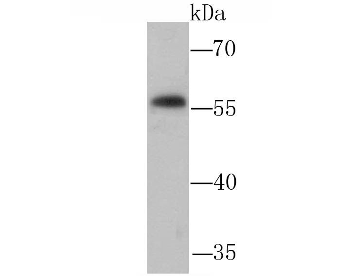
Fig1: Western blot analysis of NFIB/NF1B2 on PC-12 cell lysate using anti-NFIB/NF1B2 antibody at 1/1,000 dilution.Application
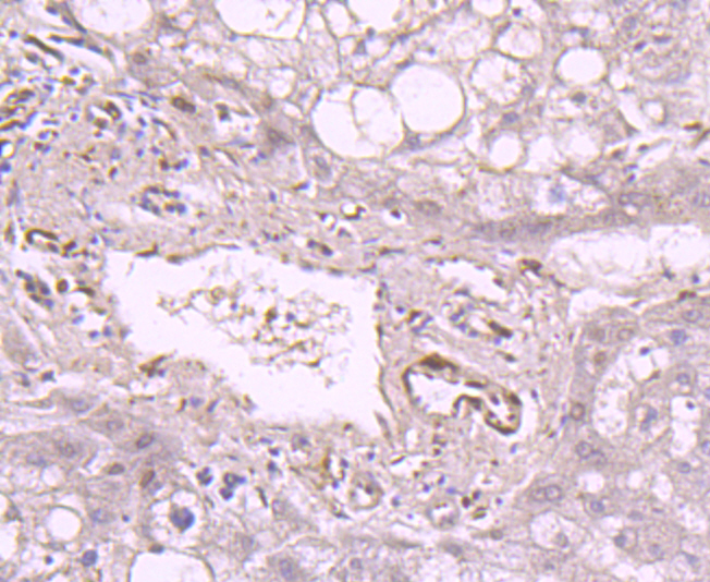
Fig2: Immunohistochemical analysis of paraffin-embedded human liver tissue using anti-NFIB/NF1B2 antibody. Counter stained with hematoxylin.Application
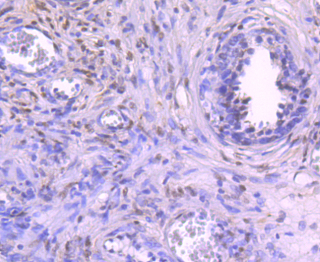
Fig3: Immunohistochemical analysis of paraffin-embedded human prostate tissue using anti-NFIB/NF1B2 antibody. Counter stained with hematoxylin.Application
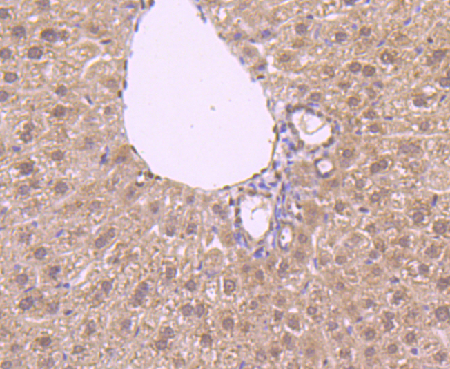
Fig4: Immunohistochemical analysis of paraffin-embedded mouse liver tissue using anti-NFIB/NF1B2 antibody. Counter stained with hematoxylin.Application
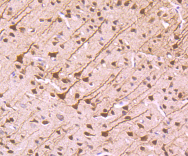
Fig5: Immunohistochemical analysis of paraffin-embedded mouse brain tissue using anti-NFIB/NF1B2 antibody. Counter stained with hematoxylin.Application
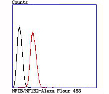
Fig6: Flow cytometric analysis of Hela cells with NFIB/NF1B2 antibody at 1/100 dilution (red) compared with an unlabelled control (cells without incubation with primary antibody; black).| Product Name | NFIB/NF1B2 |
|---|---|
| Antibody Type | Primary Antibodies |
| Antigen Alias | CCAAT Box Binding Transcription Factor antibody CCAAT-box-binding transcription factor antibody CTF antibody HMGIC/NFIB antibody NF-I/B antibody NF1-B antibody NF1B antibody NF1B2 antibody NFI-B antibody NFI-RED antibody Nfib antibody NFIB_HUMAN antibody NFIB2 antibody NFIB3 antibody Nuclear factor 1 B-type antibody Nuclear factor 1/B antibody Nuclear Factor 1B antibody Nuclear Factor I B antibody Nuclear factor I/B antibody TGGCA Binding Protein antibody TGGCA-binding protein antibody TRANSCRIPTION FACTOR NFIB antibody |
| Product description | Recognizes and binds the palindromic sequence 5'-TTGGCNNNNNGCCAA-3' present in viral and cellular promoters and in the origin of replication of adenovirus type 2. These proteins are individually capable of activating transcription and replication. |
| Immunogen | Recombinant protein |
| Clonality | Polyclonal |
|---|---|
| Isotype | IgG |
| Host Species | Rabbit |
| Tested Applications | WBIHCFC |
| WB:1:200-1:500 IHC:1:50-1:100 FC:1:50-1:100 |
|
| Species Reactivity | HumanMouse |
| Concentration | 1mg/ml |
| Alternative Names | CCAAT Box Binding Transcription Factor antibody CCAAT-box-binding transcription factor antibody CTF antibody HMGIC/NFIB antibody NF-I/B antibody NF1-B antibody NF1B antibody NF1B2 antibody NFI-B antibody NFI-RED antibody Nfib antibody NFIB_HUMAN antibody NFIB2 antibody NFIB3 antibody Nuclear factor 1 B-type antibody Nuclear factor 1/B antibody Nuclear Factor 1B antibody Nuclear Factor I B antibody Nuclear factor I/B antibody TGGCA Binding Protein antibody TGGCA-binding protein antibody TRANSCRIPTION FACTOR NFIB antibody |
|---|---|
| Molecular Weight(MW) | 56 kDa |
| Cellular Localization | Nucleus. |
Application
Fig1: Western blot analysis of NFIB/NF1B2 on PC-12 cell lysate using anti-NFIB/NF1B2 antibody at 1/1,000 dilution.
Application
Fig2: Immunohistochemical analysis of paraffin-embedded human liver tissue using anti-NFIB/NF1B2 antibody. Counter stained with hematoxylin.
Application
Fig3: Immunohistochemical analysis of paraffin-embedded human prostate tissue using anti-NFIB/NF1B2 antibody. Counter stained with hematoxylin.
Application
Fig4: Immunohistochemical analysis of paraffin-embedded mouse liver tissue using anti-NFIB/NF1B2 antibody. Counter stained with hematoxylin.
Application
Fig5: Immunohistochemical analysis of paraffin-embedded mouse brain tissue using anti-NFIB/NF1B2 antibody. Counter stained with hematoxylin.
Application
Fig6: Flow cytometric analysis of Hela cells with NFIB/NF1B2 antibody at 1/100 dilution (red) compared with an unlabelled control (cells without incubation with primary antibody; black).| Positive Control | PC-12, human liver tissue, human prostate tissue, mouse liver tissue, mouse brain tissue, Hela. |
|---|---|
| Application Notes | WB:1:200-1:500 IHC:1:50-1:100 FC:1:50-1:100 |
| Form | Liquid |
|---|---|
| Storage Instructions | Store at +4℃ after thawing. Aliquot store at -20℃ or -80℃. Avoid repeated freeze / thaw cycles. |
| Storage Buffer | 1*TBS (pH7.4), 0.5%BSA, 50%Glycerol. Preservative: 0.05% Sodium Azide. |


