Application
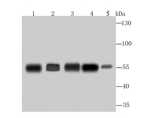
Fig1: Western blot analysis of G6PD on different lysates using anti-G6PD antibody at 1/1,000 dilution. Positive control:Lane1: A549Lane2: HelaLane3: MCF-7Lane4: PC-12Lane 5: Mouse spleenApplication
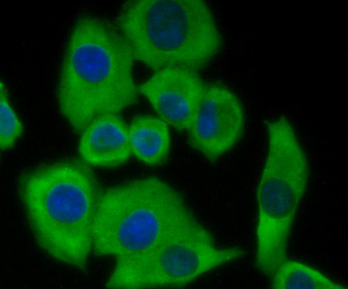
Fig2: ICC staining G6PD in A549 cells (green). The nuclear counter stain is DAPI (blue). Cells were fixed in paraformaldehyde, permeabilised with 0.25% Triton X100/PBS.Application
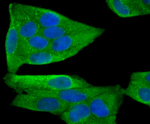
Fig3: ICC staining G6PD in Hela cells (green). The nuclear counter stain is DAPI (blue). Cells were fixed in paraformaldehyde, permeabilised with 0.25% Triton X100/PBS.Application
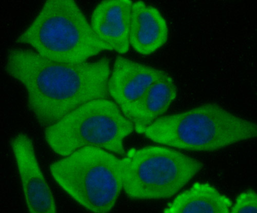
Fig4: ICC staining G6PD in MCF-7 cells (green). The nuclear counter stain is DAPI (blue). Cells were fixed in paraformaldehyde, permeabilised with 0.25% Triton X100/PBS.Application
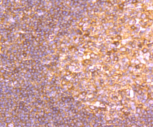
Fig5: Immunohistochemical analysis of paraffin-embedded human tonsil tissue using anti-G6PD antibody. Counter stained with hematoxylin.Application
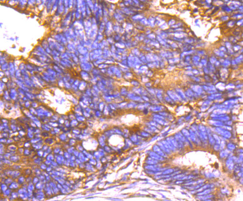
Fig6: Immunohistochemical analysis of paraffin-embedded human colon cancer tissue using anti-G6PD antibody. Counter stained with hematoxylin.Application
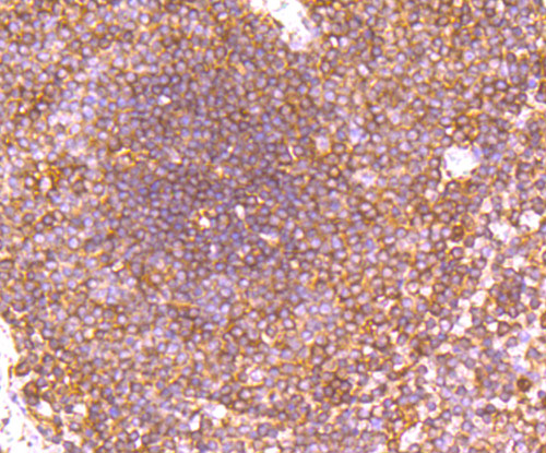
Fig7: Immunohistochemical analysis of paraffin-embedded human spleen tissue using anti-G6PD antibody. Counter stained with hematoxylin.Application
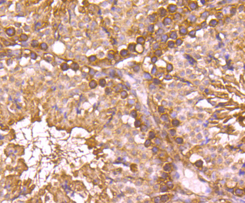
Fig8: Immunohistochemical analysis of paraffin-embedded mouse testis tissue using anti-G6PD antibody. Counter stained with hematoxylin.| Product Name | G6PD |
|---|---|
| Antibody Type | Primary Antibodies |
| Antigen Alias | G6PD antibody G6PD_HUMAN antibody G6PD1 antibody G6pdx antibody Glucose 6 phosphate 1 dehydrogenase antibody Glucose 6 phosphate dehydrogenase antibody Glucose 6 phosphate dehydrogenase, G6PD antibody Glucose-6-phosphate 1-dehydrogenase antibody MET19 antibody POS10 antibody Zwf1p antibody |
| Product description | Glucose-6-phosphate 1-dehydrogenase (G6PD) plays an important role in the pentose phosphate pathway. It is a member of the glucose-6-phosphate dehydrogenase family of proteins. G6PD is a ubiquitous enzyme that produces pentose sugars for nucleic acid synthesis, but is also involved in carbohydrate degradation, as it is one of the main producers of NADPH reducing power. G6PD has NADP as a co-factor and structural element. It can be found as a homodimer or homotetramer, and is primarily detected in lymphoblasts, granulocytes and sperm. Defects in G6PD can cause chronic non-spherocytic hemolytic anemia (CNSHA), especially in areas in which malaria is an epidemic. Individuals with a high level of G6PD-deficiency are at higher risk of acute hemolytic attacks. |
| Immunogen | Recombinant protein. |
| Clonality | Polyclonal |
|---|---|
| Isotype | IgG |
| Host Species | Rabbit |
| Tested Applications | WBICCIHC |
| WB:1:500~1:2000 ICC:1:200~1:500 IHC:1:50~1:200 Notes:Optimal dilutions/concentrations should be determined by the researcher. |
|
| Species Reactivity | HumanMouseRat |
| Concentration | 1mg/ml |
| Alternative Names | G6PD antibody
G6PD_HUMAN antibody
G6PD1 antibody
G6pdx antibody
Glucose 6 phosphate 1 dehydrogenase antibody
Glucose 6 phosphate dehydrogenase antibody
Glucose 6 phosphate dehydrogenase G6PD antibody Glucose-6-phosphate 1-dehydrogenase antibody MET19 antibody POS10 antibody Zwf1p antibody |
|---|---|
| Molecular Weight(MW) | 59 kDa |
| Cellular Localization | Cytoplasm. Nucleus. Membrane. |
Application
Fig1: Western blot analysis of G6PD on different lysates using anti-G6PD antibody at 1/1,000 dilution. Positive control:Lane1: A549Lane2: HelaLane3: MCF-7Lane4: PC-12Lane 5: Mouse spleen
Application
Fig2: ICC staining G6PD in A549 cells (green). The nuclear counter stain is DAPI (blue). Cells were fixed in paraformaldehyde, permeabilised with 0.25% Triton X100/PBS.
Application
Fig3: ICC staining G6PD in Hela cells (green). The nuclear counter stain is DAPI (blue). Cells were fixed in paraformaldehyde, permeabilised with 0.25% Triton X100/PBS.
Application
Fig4: ICC staining G6PD in MCF-7 cells (green). The nuclear counter stain is DAPI (blue). Cells were fixed in paraformaldehyde, permeabilised with 0.25% Triton X100/PBS.
Application
Fig5: Immunohistochemical analysis of paraffin-embedded human tonsil tissue using anti-G6PD antibody. Counter stained with hematoxylin.
Application
Fig6: Immunohistochemical analysis of paraffin-embedded human colon cancer tissue using anti-G6PD antibody. Counter stained with hematoxylin.
Application
Fig7: Immunohistochemical analysis of paraffin-embedded human spleen tissue using anti-G6PD antibody. Counter stained with hematoxylin.
Application
Fig8: Immunohistochemical analysis of paraffin-embedded mouse testis tissue using anti-G6PD antibody. Counter stained with hematoxylin.| Positive Control | A549, Hela, MCF-7, PC-12, mouse spleen tissue lysate, human tonsil tissue, human colon cancer tissue, human spleen tissue, mouse testis tissue. |
|---|---|
| Application Notes | WB:1:500~1:2000 ICC:1:200~1:500 IHC:1:50~1:200 Notes:Optimal dilutions/concentrations should be determined by the researcher. |
| Form | Liquid |
|---|---|
| Storage Instructions | Store at +4℃ after thawing. Aliquot store at -20℃ or -80℃. Avoid repeated freeze / thaw cycles. |
| Storage Buffer | 1*TBS (pH7.4), 0.5%BSA, 50%Glycerol. Preservative: 0.05% Sodium Azide. |


