Application
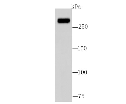
Fig1: Western blot analysis of Fatty Acid Synthase on A549 cell lysate using anti-Fatty Acid Synthase antibody at 1/1,000 dilution.Application
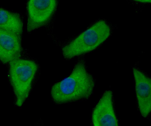
Fig2: ICC staining Fatty Acid Synthase in A549 cells (green). The nuclear counter stain is DAPI (blue). Cells were fixed in paraformaldehyde, permeabilised with 0.25% Triton X100/PBS.Application
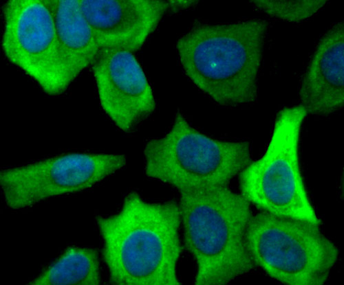
Fig3: ICC staining Fatty Acid Synthase in MCF-7 cells (green). The nuclear counter stain is DAPI (blue). Cells were fixed in paraformaldehyde, permeabilised with 0.25% Triton X100/PBS.Application
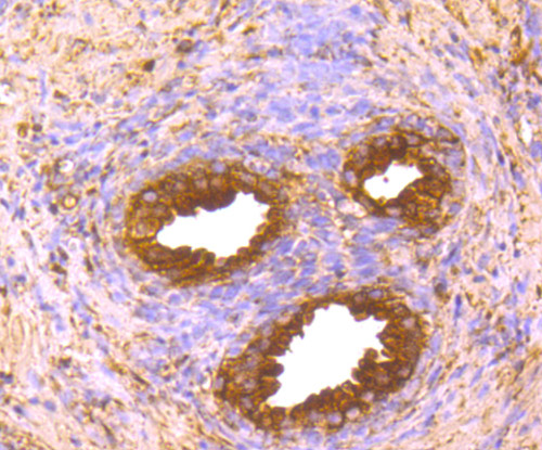
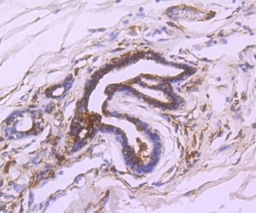
Fig4: Immunohistochemical analysis of paraffin-embedded human prostate tissue using anti-Fatty Acid Synthase antibody. Counter stained with hematoxylin.Application
Fig5: Immunohistochemical analysis of paraffin-embedded human breast cancer tissue using anti-Fatty Acid Synthase antibody. Counter stained with hematoxylin.| Product Name | Fatty Acid Synthase |
|---|---|
| Antibody Type | Primary Antibodies |
| Antigen Alias | [Acyl-carrier-protein] S acetyltransferase antibody [Acyl-carrier-protein] S malonyltransferase antibody 3-hydroxypalmitoyl-[acyl-carrier-protein] dehydratase antibody 3-oxoacyl-[acyl-carrier-protein] reductase antibody 3-oxoacyl-[acyl-carrier-protein] synthase antibody Enoyl-[acyl-carrier-protein] reductase antibody FAS antibody FAS_HUMAN antibody FASN antibody Fatty acid synthase antibody MGC14367 antibody MGC15706 antibody OA 519 antibody Oleoyl-[acyl-carrier-protein] hydrolase antibody SDR27X1 antibody Short chain dehydrogenase/reductase family 27X member 1 antibody |
| Product description | Fatty acid biosynthesis is mediated by seven catalytic enzymes and an acyl carrier protein (ACP), to which various acyl intermediates are covalently attached. Fatty Acid Synthase (FAS) is the anabolic enzyme that contains the seven unique catalytic sites and mediates the conversion of acetyl-CoA and malonyl-CoA, in the presence of the cofactor NADPH, into long-chain saturated fatty acids, such as palmitate. Human Fatty Acid Synthase cDNA encodes a 2,504 amino acid protein. Catalytically active Fatty Acid Synthase is a homodimer. Human Fatty Acid Synthase mRNA is variably expressed with abundant levels present in brain, lung and liver. Fatty acid synthetic metabolism is abnormally elevated in tumor cells and may support cell growth or survival of malignant cancers. |
| Immunogen | Recombinant protein. |
| Clonality | Polyclonal |
|---|---|
| Isotype | IgG |
| Host Species | Rabbit |
| Tested Applications | WBICCIHC |
| WB:1:500~1:2000 ICC:1:200~1:500 IHC:1:50~1:200 Notes:Optimal dilutions/concentrations should be determined by the researcher. |
|
| Species Reactivity | HumanMouseRat |
| Concentration | 1mg/ml |
| Alternative Names | [Acyl-carrier-protein] S acetyltransferase antibody [Acyl-carrier-protein] S malonyltransferase antibody 3-hydroxypalmitoyl-[acyl-carrier-protein] dehydratase antibody 3-oxoacyl-[acyl-carrier-protein] reductase antibody 3-oxoacyl-[acyl-carrier-protein] synthase antibody Enoyl-[acyl-carrier-protein] reductase antibody FAS antibody FAS_HUMAN antibody FASN antibody Fatty acid synthase antibody MGC14367 antibody MGC15706 antibody OA 519 antibody Oleoyl-[acyl-carrier-protein] hydrolase antibody SDR27X1 antibody Short chain dehydrogenase/reductase family 27X member 1 antibody |
|---|---|
| Molecular Weight(MW) | 273 kDa |
| Cellular Localization | Cytoplasm. Melanosome. |
Application
Fig1: Western blot analysis of Fatty Acid Synthase on A549 cell lysate using anti-Fatty Acid Synthase antibody at 1/1,000 dilution.
Application
Fig2: ICC staining Fatty Acid Synthase in A549 cells (green). The nuclear counter stain is DAPI (blue). Cells were fixed in paraformaldehyde, permeabilised with 0.25% Triton X100/PBS.
Application
Fig3: ICC staining Fatty Acid Synthase in MCF-7 cells (green). The nuclear counter stain is DAPI (blue). Cells were fixed in paraformaldehyde, permeabilised with 0.25% Triton X100/PBS.
Application
Fig4: Immunohistochemical analysis of paraffin-embedded human prostate tissue using anti-Fatty Acid Synthase antibody. Counter stained with hematoxylin.
Application
Fig5: Immunohistochemical analysis of paraffin-embedded human breast cancer tissue using anti-Fatty Acid Synthase antibody. Counter stained with hematoxylin.| Positive Control | A549, MCF-7, human prostate tissue, human breast cancer tissue. |
|---|---|
| Application Notes | WB:1:500~1:2000 ICC:1:200~1:500 IHC:1:50~1:200 Notes:Optimal dilutions/concentrations should be determined by the researcher. |
| Form | Liquid |
|---|---|
| Storage Instructions | Store at +4℃ after thawing. Aliquot store at -20℃ or -80℃. Avoid repeated freeze / thaw cycles. |
| Storage Buffer | 1*TBS (pH7.4), 0.5%BSA, 50%Glycerol. Preservative: 0.05% Sodium Azide. |


