Application
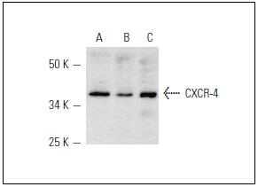
Fig1: Western blot analysis of CXCR-4 expression in HL-60 (A), U-937 (B) and Jurkat (C) whole cell lysates.Application
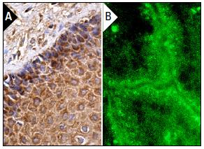
Fig2: Immunoperoxidase staining of formalin fixed, paraffin-embedded human cervix tissue showing cytoplasmic staining of squamous epithelial cells (A). Immunofluorescence staining of normal mouse intestine frozen section showing membrane staining (B).| Product Name | CXCR4 |
|---|---|
| Antibody Type | Primary Antibodies |
| Antigen Alias | C-X-C chemokine receptor type 4 antibody CD184 antibody CD184 antigen antibody Chemokine (C X C motif) receptor 4 antibody Chemokine CXC Motif Receptor 4 antibody CXC-R4 antibody CXCR-4 antibody CXCR4 antibody CXCR4_HUMAN antibody D2S201E antibody FB22 antibody Fusin antibody HM89 antibody HSY3RR antibody LAP 3 antibody LAP3 antibody LCR1 antibody LESTR antibody Leukocyte derived seven transmembrane domain receptor antibody Leukocyte-derived seven transmembrane domain receptor antibody Lipopolysaccharide associated protein 3 antibody Neuropeptide Y receptor Y3 antibody NPY3R antibody NPYR antibody NPYRL antibody NPYY3 antibody NPYY3R antibody Probable G protein coupled receptor lcr1 homolog antibody SDF 1 receptor antibody SDF-1 receptor antibody SEVEN-TRANSMEMBRANE-SEGMENT RECEPTOR antibody Stromal cell derived factor 1 receptor antibody Stromal cell-derived factor 1 receptor antibody WHIM antibody WHIMS antibody |
| Product description | Cell membrane, Cell junction, Lysosome, Early/ Late endosome |
| Immunogen | Amino acids 176-293 of CXCR-4 of human origin. |
| Clonality | Polyclonal |
|---|---|
| Isotype | IgG |
| Host Species | Rabbit |
| Tested Applications | WB |
| WB:1:100-1:1,000 |
|
| Species Reactivity | HumanMouseRat |
| Concentration | 1mg/ml |
| Alternative Names | C-X-C chemokine receptor type 4 antibody CD184 antibody CD184 antigen antibody Chemokine (C X C motif) receptor 4 antibody Chemokine CXC Motif Receptor 4 antibody CXC-R4 antibody CXCR-4 antibody CXCR4 antibody CXCR4_HUMAN antibody D2S201E antibody FB22 antibody Fusin antibody HM89 antibody HSY3RR antibody LAP 3 antibody LAP3 antibody LCR1 antibody LESTR antibody Leukocyte derived seven transmembrane domain receptor antibody Leukocyte-derived seven transmembrane domain receptor antibody Lipopolysaccharide associated protein 3 antibody Neuropeptide Y receptor Y3 antibody NPY3R antibody NPYR antibody NPYRL antibody NPYY3 antibody NPYY3R antibody Probable G protein coupled receptor lcr1 homolog antibody SDF 1 receptor antibody SDF-1 receptor antibody SEVEN-TRANSMEMBRANE-SEGMENT RECEPTOR antibody Stromal cell derived factor 1 receptor antibody Stromal cell-derived factor 1 receptor antibody WHIM antibody WHIMS antibody |
|---|---|
| Molecular Weight(MW) | 40-47kDa |
| Cellular Localization | Cell membrane, Cell junction, Lysosome, |
| SwissProt ID | P61073 |
|---|
Application
Fig1: Western blot analysis of CXCR-4 expression in HL-60 (A), U-937 (B) and Jurkat (C) whole cell lysates.
Application
Fig2: Immunoperoxidase staining of formalin fixed, paraffin-embedded human cervix tissue showing cytoplasmic staining of squamous epithelial cells (A). Immunofluorescence staining of normal mouse intestine frozen section showing membrane staining (B).| Positive Control | HL-60, U-937, Jurkat, human cervix tissue. |
|---|---|
| Application Notes | WB:1:100-1:1,000 |
| Form | Liquid |
|---|---|
| Storage Instructions | Store at +4℃ |
| Storage Buffer | 1*TBS (pH7.4), 1%BSA, 40%Glycerol. Preservative: 0.05% Sodium Azide. |


