Application
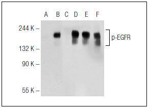
Fig1: Western blot analysis of EGFR phosphorylation in untreated (A, D), EGF treated (B, E) and EGF and lambda protein phosphatase treated (C, F) A-431 whole cell lysates. Antibodies tested include p-EGFR (Tyr 1173) (A, B, C) and EGFR (1005) (D, E, F).Application
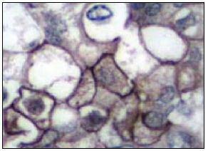
Fig2: Immunoperoxidase staining of formalin-fixed, paraffin-embedded human breast carcinoma tissue showing membrane staining.| Product Name | Phospho-EGFR (Tyr 1173) |
|---|---|
| Antibody Type | Primary Antibodies |
| Antigen Alias | Avian erythroblastic leukemia viral (v erb b) oncogene homolog antibody Cell growth inhibiting protein 40 antibody Cell proliferation inducing protein 61 antibody EGF R antibody EGFR antibody EGFR_HUMAN antibody Epidermal growth factor receptor (avian erythroblastic leukemia viral (v erb b) oncogene homolog) antibody Epidermal growth factor receptor (erythroblastic leukemia viral (v erb b) oncogene homolog avian) antibody Epidermal growth factor receptor antibody erb-b2 receptor tyrosine kinase 1 antibody ERBB antibody ERBB1 antibody Errp antibody HER1 antibody mENA antibody NISBD2 antibody Oncogen ERBB antibody PIG61 antibody Proto-oncogene c-ErbB-1 antibody Receptor tyrosine protein kinase ErbB 1 antibody Receptor tyrosine-protein kinase ErbB-1 antibody SA7 antibody Species antigen 7 antibody Urogastrone antibody v-erb-b Avian erythroblastic leukemia viral oncogen homolog antibody wa2 antibody Wa5 antibody hide |
| Product description | Epidermal growth factor mediates its effects on cell growth through its inter-action with a cell surface glycoprotein designated the EGF receptor. Binding of EGF or TGF alpha to the EGF receptor activates tyrosine-specific protein kinase activity intrinsic to the EGF receptor. The carboxy terminal tyrosine residues on EGFR, Tyr 1068 and Tyr 1173, are the major sites of autophosphorylation, which occurs as a result of EGF binding. Once activated, EGFR mediates the binding of the phosphotyrosine binding (PTB) domain of GRB2 through direct interactions with Tyr 1068 and Tyr 1086 and through indirect interactions with Tyr 1173 in the Ras signaling pathway. Tyr 1173 of EGFR also functions as a kinase substrate. Phosphorylation of Tyr 992, Tyr 1068 and Tyr 1086 is required for conformational change in the C-terminal tail of the EGF receptor. |
| Immunogen | A short amino acid sequence containing phosphorylated Tyr 1173 of EGFR of human origin. |
| Modification | p-Tyr 1173 |
| Clonality | Polyclonal |
|---|---|
| Isotype | IgG |
| Host Species | Rabbit |
| Tested Applications | WBIPIFIHC-P |
| WB:1:100-1:1,000 IP:1-2 μg per 100-500 μg of total protein (1 ml of cell lysate) IF:1:50-1:500 IHC:1:50-1:500 |
|
| Species Reactivity | HumanMouseRat |
| Concentration | 1mg/ml |
| Alternative Names | Avian erythroblastic leukemia viral (v erb b) oncogene homolog antibody Cell growth inhibiting protein 40 antibody Cell proliferation inducing protein 61 antibody EGF R antibody EGFR antibody EGFR_HUMAN antibody Epidermal growth factor receptor (avian erythroblastic leukemia viral (v erb b) oncogene homolog) antibody Epidermal growth factor receptor (erythroblastic leukemia viral (v erb b) oncogene homolog avian) antibody Epidermal growth factor receptor antibody erb-b2 receptor tyrosine kinase 1 antibody ERBB antibody ERBB1 antibody Errp antibody HER1 antibody mENA antibody NISBD2 antibody Oncogen ERBB antibody PIG61 antibody Proto-oncogene c-ErbB-1 antibody Receptor tyrosine protein kinase ErbB 1 antibody Receptor tyrosine-protein kinase ErbB-1 antibody SA7 antibody Species antigen 7 antibody Urogastrone antibody v-erb-b Avian erythroblastic leukemia viral oncogen homolog antibody wa2 antibody Wa5 antibody hide |
|---|---|
| Molecular Weight(MW) | 170 kDa |
| Cellular Localization | Cell membrane, Secreted, Endosome, Nucleus, Endosome, Nucleus membrane, Endosome membrane |
| SwissProt ID | P00533 |
|---|
Application
Fig1: Western blot analysis of EGFR phosphorylation in untreated (A, D), EGF treated (B, E) and EGF and lambda protein phosphatase treated (C, F) A-431 whole cell lysates. Antibodies tested include p-EGFR (Tyr 1173) (A, B, C) and EGFR (1005) (D, E, F).
Application
Fig2: Immunoperoxidase staining of formalin-fixed, paraffin-embedded human breast carcinoma tissue showing membrane staining.| Positive Control | A-431, human breast carcinoma tissue. |
|---|---|
| Application Notes | WB:1:100-1:1,000 IP:1-2 μg per 100-500 μg of total protein (1 ml of cell lysate) IF:1:50-1:500 IHC:1:50-1:500 |
| Form | Liquid |
|---|---|
| Storage Instructions | Store at +4℃ |
| Storage Buffer | 1*TBS (pH7.4), 1%BSA, 40%Glycerol. Preservative: 0.05% Sodium Azide. |


