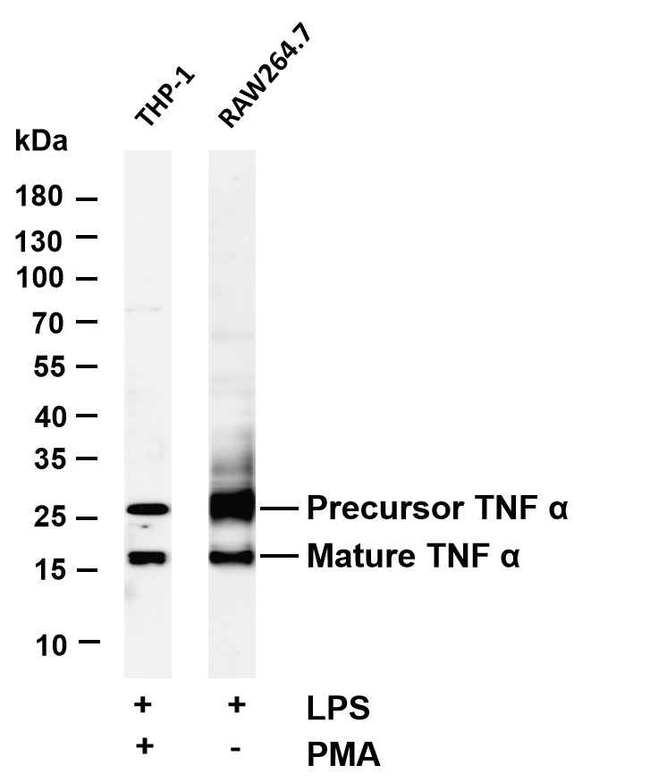

WB
Various whole cell lysates were separated by 4-20% SDS-PAGE, and the membrane was blotted with anti-protein name antibody. The HRP-conjugated Goat anti-Rabbit IgG(H + L) antibody was used to detect the antibody. Lane 1:THP-1 tearted with Phorbol 12-myristate 13-acetate and Lipopolysaccharide (100 mg/mL) for 24 hours. Lane 2: RAW264.7 tearted with Lipopolysaccharide (100 mg/mL) for 24 hours .Predicted band size: 26kDa Observed band size: 26kDa.IHC
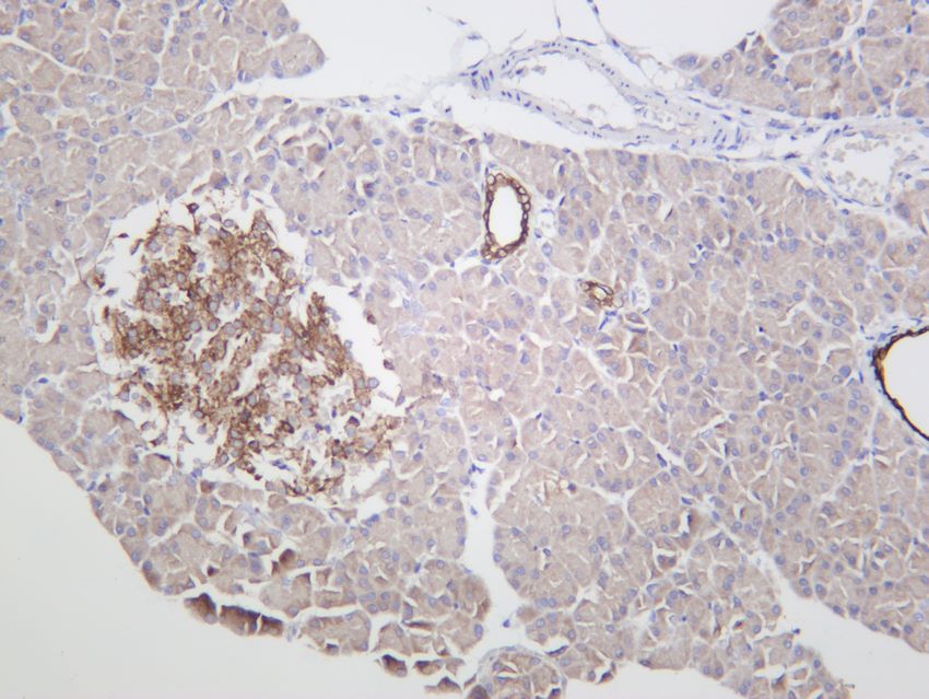
Rat pancreas was stained with anti-TNFα rabbit antibody.ICC/IF
Immunofluorescence analysis of A549. 1,primary Antibody(red) was diluted at 1:200(4°C overnight). 2, Goat Anti Rabbit IgG (H&L) - Alexa Fluor 594 Secondary antibody was diluted at 1:1000(room temperature, 50min).3,DAPI(blue) 10min.| Product Name | TNFα Rabbit mAb |
|---|---|
| Antibody Type | Primary Antibodies |
| Clonality | monoclonal |
|---|---|
| Isotype | IgG |
| Host Species | Rabbit |
| Tested Applications | ICC/IFIHCWB |
| WB:1:2000-1:10000 IHC:1:2000-1:10000 ICC/IF:1:200-1:1000 |
|
| Species Reactivity | HumanMouseRat |
| Concentration | 1mg/ml |
| Purification | Protein A |
| Gene Symbol | TNF |
|---|---|
| Gene Synonyms | DIF TNFA IMD127 TNFSF2 TNLG1F TNF-alpha |
| Gene Full Name | tumor necrosis factor |
| Gene Summary | This gene encodes a multifunctional proinflammatory cytokine that belongs to the tumor necrosis factor (TNF) superfamily. This cytokine is mainly secreted by macrophages. It can bind to, and thus functions through its receptors TNFRSF1A/TNFR1 and TNFRSF1B/TNFBR. This cytokine is involved in the regulation of a wide spectrum of biological processes including cell proliferation, differentiation, apoptosis, lipid metabolism, and coagulation. This cytokine has been implicated in a variety of diseases, including autoimmune diseases, insulin resistance, psoriasis, rheumatoid arthritis ankylosing spondylitis, tuberculosis, autosomal dominant polycystic kidney disease, and cancer. Mutations in this gene affect susceptibility to cerebral malaria, septic shock, and Alzheimer disease. Knockout studies in mice also suggested the neuroprotective function of this cytokine. [provided by RefSeq, Aug 2020] |
| Molecular Weight(MW) | 18kDa |
| Cellular Localization | Membrane. |
WB
Various whole cell lysates were separated by 4-20% SDS-PAGE, and the membrane was blotted with anti-protein name antibody. The HRP-conjugated Goat anti-Rabbit IgG(H + L) antibody was used to detect the antibody. Lane 1:THP-1 tearted with Phorbol 12-myristate 13-acetate and Lipopolysaccharide (100 mg/mL) for 24 hours. Lane 2: RAW264.7 tearted with Lipopolysaccharide (100 mg/mL) for 24 hours .Predicted band size: 26kDa Observed band size: 26kDa.
IHC
Rat pancreas was stained with anti-TNFα rabbit antibody.
ICC/IF
Immunofluorescence analysis of A549. 1,primary Antibody(red) was diluted at 1:200(4°C overnight). 2, Goat Anti Rabbit IgG (H&L) - Alexa Fluor 594 Secondary antibody was diluted at 1:1000(room temperature, 50min).3,DAPI(blue) 10min.| Application Notes | WB:1:2000-1:10000 IHC:1:2000-1:10000 ICC/IF:1:200-1:1000 |
|---|
| Form | Liquid |
|---|---|
| Storage Instructions | -15°C to -25°C/1 year(Do not lower than -25°C) |
| Storage Buffer | PBS, 50% glycerol, 0.05% Proclin 300, 0.05%BSA |


 Data sheet for OM643883