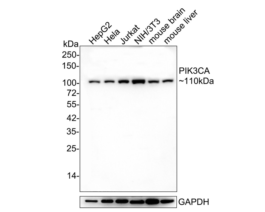
WB
Western blot analysis of PI3 Kinase p110α on different lysates with Rabbit anti-PI3 Kinase p110α antibody at 1/1,000 dilution. Lane 1: HepG2 cell lysate, Lane 2: Hela cell lysate, Lane 3: Jurkat cell lysate, Lane 4: NIH/3T3 cell lysate, Lane 5: mouse brain tissue lysate(20 µg/Lane), Lane 6: mouse liver tissue lysate(20 µg/Lane), Lysates/proteins at 10 µg/Lane. Exposure time: 3 minutes; 4-20% SDS-PAGE gel. Proteins were transferred to a PVDF membrane and blocked with 5% NFDM/TBST for 1 hour at room temperature. The primary antibody at 1/1,000 dilution was used in 5% NFDM/TBST at 4℃ overnight. Goat Anti-Rabbit IgG - HRP Secondary Antibody at 1/50,000 dilution was used for 1 hour at room temperature.IHC
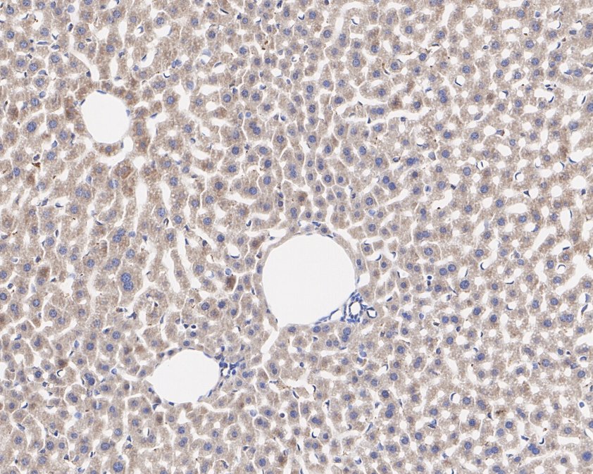
Immunohistochemical analysis of paraffin-embedded mouse liver tissue with Rabbit anti-PI3 Kinase p110α antibody at 1/200 dilution. The section was pre-treated using heat mediated antigen retrieval with Tris-EDTA buffer (pH 9.0) for 20 minutes. The tissues were blocked in 1% BSA for 20 minutes at room temperature, washed with ddH2O and PBS, and then probed with the primary antibody at 1/200 dilution for 1 hour at room temperature. The detection was performed using an HRP conjugated compact polymer system. DAB was used as the chromogen. Tissues were counterstained with hematoxylin and mounted with DPX.ICC/IF
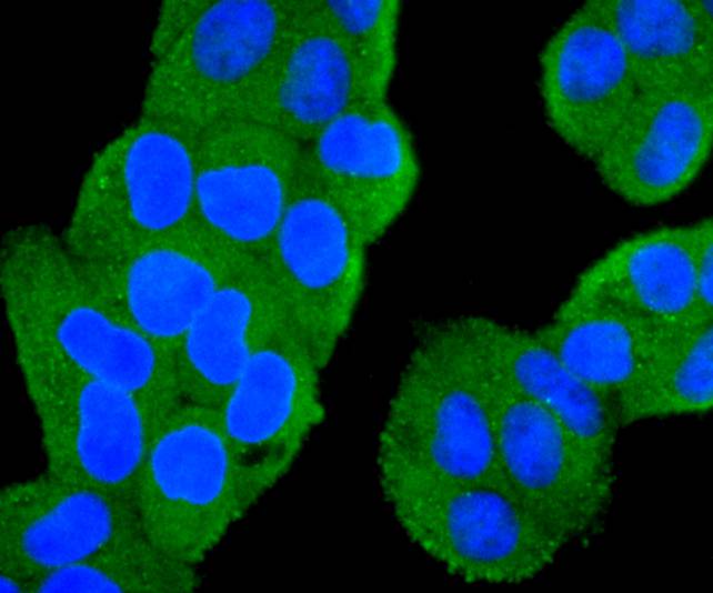
ICC staining of PI3 Kinase p110α in Hela cells (green). Formalin fixed cells were permeabilized with 0.1% Triton X-100 in TBS for 10 minutes at room temperature and blocked with 1% Blocker BSA for 15 minutes at room temperature. Cells were probed with the primary antibody (1/50) for 1 hour at room temperature, washed with PBS. Alexa Fluor®488 Goat anti-Rabbit IgG was used as the secondary antibody at 1/1,000 dilution. The nuclear counter stain is DAPI (blue).IP
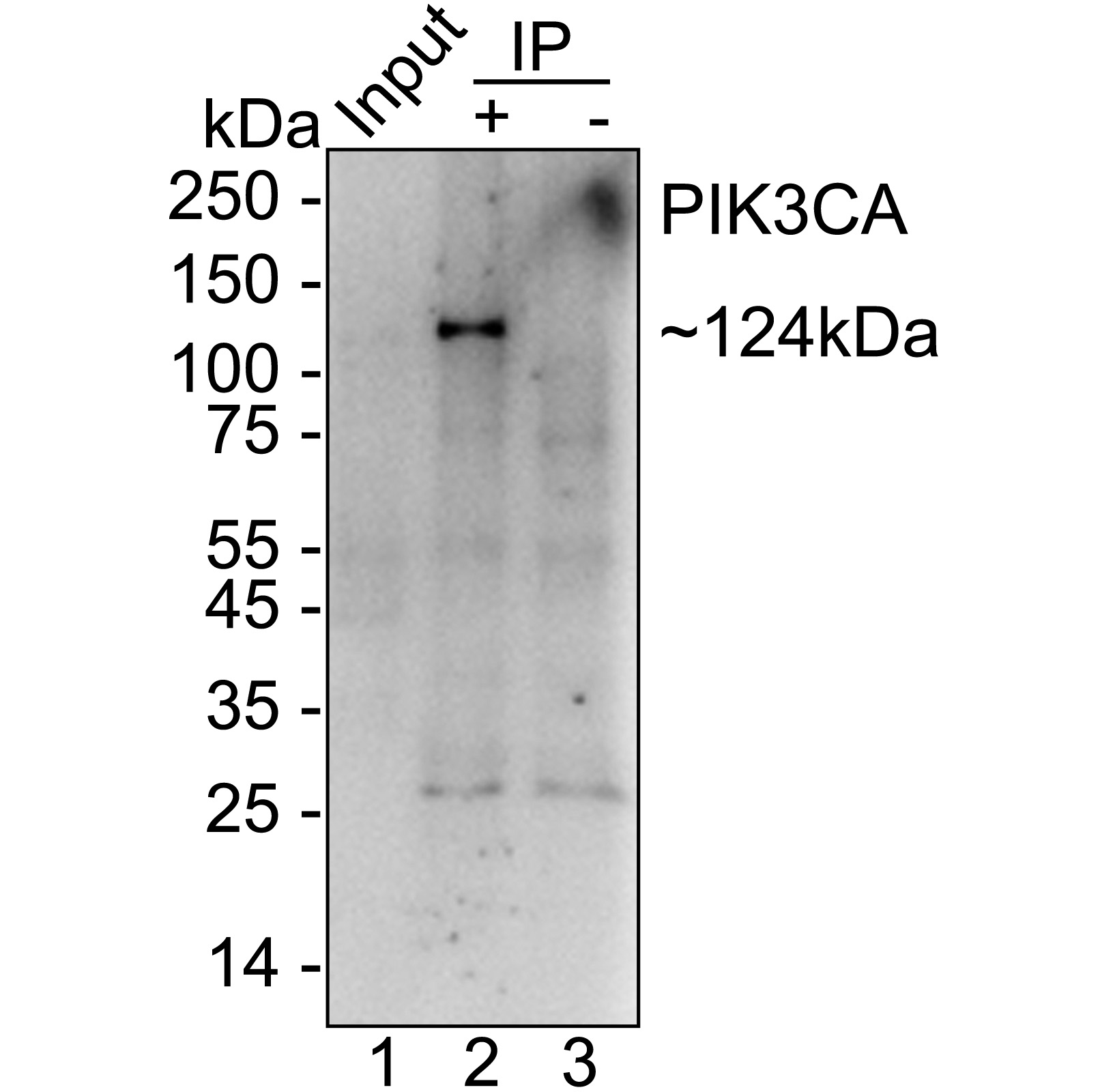
PI3 Kinase p110α was immunoprecipitated from 0.2 mg HeLa cell lysate with with Rabbit anti-PI3 Kinase p110α antibody at 2 µg/25 µl agarose. Western blot was performed from the immunoprecipitate usingwith Rabbit anti-PI3 Kinase p110α antibody at 1/1,000 dilution. Anti-Rabbit IgG for IP Nano-secondary antibody at 1/5,000 dilution was used for 1 hour at room temperature. Lane 1: HeLa cell lysate (input), Lane 2: with Rabbit anti-PI3 Kinase p110α antibody IP in HeLa cell lysate, Lane 3: Rabbit IgG instead ofwith Rabbit anti-PI3 Kinase p110α antibody in HeLa cell lysate, Blocking/Dilution buffer: 5% NFDM/TBST, Exposure time: 2 minutes.| Product Name | PI3 Kinase p110α Recombinant Rabbit Monoclonal Antibody |
|---|---|
| Antibody Type | Primary Antibodies |
| Immunogen | Synthetic peptide within Human PIK3CA aa 1,001-1,050 / 1,068. |
| Clonality | monoclonal |
|---|---|
| Isotype | IgG |
| Host Species | Rabbit |
| Tested Applications | ICC/IFIHCIPWB |
| WB:1:1000 IHC:1:50-1:200 ICC/IF:1:50-1:200 IP:1-2μg/sample |
|
| Species Reactivity | HumanMouse |
| Concentration | 1mg/ml |
| Purification | Protein A |
| Gene Symbol | PIK3CA |
|---|---|
| Gene Synonyms | HMH MCM CCM4 CWS5 MCAP PI3K CLAPO CLOVE MCMTC PI3K-alpha p110-alpha |
| Gene Full Name | phosphatidylinositol-4,5-bisphosphate 3-kinase catalytic subunit alpha |
| Gene Summary | Phosphatidylinositol 3-kinase is composed of an 85 kDa regulatory subunit and a 110 kDa catalytic subunit. The protein encoded by this gene represents the catalytic subunit, which uses ATP to phosphorylate PtdIns, PtdIns4P and PtdIns(4,5)P2. This gene has been found to be oncogenic and has been implicated in cervical cancers. A pseudogene of this gene has been defined on chromosome 22. [provided by RefSeq, Apr 2016] |
| Molecular Weight(MW) | 124kDa(Observed band size: 110kDa) |
| Cellular Localization | Cytosol, plasma membrane, cytoplasm, lamellipodium, membrane, phosphatidylinositol 3-kinase complex, phosphatidylinositol 3-kinase complex, class IA. |
WB
Western blot analysis of PI3 Kinase p110α on different lysates with Rabbit anti-PI3 Kinase p110α antibody at 1/1,000 dilution. Lane 1: HepG2 cell lysate, Lane 2: Hela cell lysate, Lane 3: Jurkat cell lysate, Lane 4: NIH/3T3 cell lysate, Lane 5: mouse brain tissue lysate(20 µg/Lane), Lane 6: mouse liver tissue lysate(20 µg/Lane), Lysates/proteins at 10 µg/Lane. Exposure time: 3 minutes; 4-20% SDS-PAGE gel. Proteins were transferred to a PVDF membrane and blocked with 5% NFDM/TBST for 1 hour at room temperature. The primary antibody at 1/1,000 dilution was used in 5% NFDM/TBST at 4℃ overnight. Goat Anti-Rabbit IgG - HRP Secondary Antibody at 1/50,000 dilution was used for 1 hour at room temperature.
IHC
Immunohistochemical analysis of paraffin-embedded mouse liver tissue with Rabbit anti-PI3 Kinase p110α antibody at 1/200 dilution. The section was pre-treated using heat mediated antigen retrieval with Tris-EDTA buffer (pH 9.0) for 20 minutes. The tissues were blocked in 1% BSA for 20 minutes at room temperature, washed with ddH2O and PBS, and then probed with the primary antibody at 1/200 dilution for 1 hour at room temperature. The detection was performed using an HRP conjugated compact polymer system. DAB was used as the chromogen. Tissues were counterstained with hematoxylin and mounted with DPX.
ICC/IF
ICC staining of PI3 Kinase p110α in Hela cells (green). Formalin fixed cells were permeabilized with 0.1% Triton X-100 in TBS for 10 minutes at room temperature and blocked with 1% Blocker BSA for 15 minutes at room temperature. Cells were probed with the primary antibody (1/50) for 1 hour at room temperature, washed with PBS. Alexa Fluor®488 Goat anti-Rabbit IgG was used as the secondary antibody at 1/1,000 dilution. The nuclear counter stain is DAPI (blue).
IP
PI3 Kinase p110α was immunoprecipitated from 0.2 mg HeLa cell lysate with with Rabbit anti-PI3 Kinase p110α antibody at 2 µg/25 µl agarose. Western blot was performed from the immunoprecipitate usingwith Rabbit anti-PI3 Kinase p110α antibody at 1/1,000 dilution. Anti-Rabbit IgG for IP Nano-secondary antibody at 1/5,000 dilution was used for 1 hour at room temperature. Lane 1: HeLa cell lysate (input), Lane 2: with Rabbit anti-PI3 Kinase p110α antibody IP in HeLa cell lysate, Lane 3: Rabbit IgG instead ofwith Rabbit anti-PI3 Kinase p110α antibody in HeLa cell lysate, Blocking/Dilution buffer: 5% NFDM/TBST, Exposure time: 2 minutes.| Application Notes | WB:1:1000 IHC:1:50-1:200 ICC/IF:1:50-1:200 IP:1-2μg/sample |
|---|
| Form | Liquid |
|---|---|
| Storage Instructions | Store at +4℃ after thawing. Aliquot store at -20℃. Avoid repeated freeze / thaw cycles. |
| Storage Buffer | 1*TBS (pH7.4), 0.05% BSA, 40% Glycerol. Preservative: 0.05% Sodium Azide. |


 Data sheet for OM644122