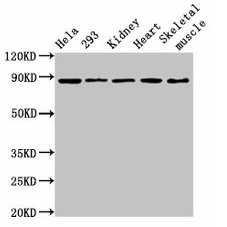
WB
Western Blot Positive WB detected in: Hela whole cell lysate, 293 whole cell lysate, Rat kidney tissue, Mouse heart tissue, Mouse skeletal muscle tissue All lanes: SEMA3A antibody at 3.6µg/ml Secondary Goat polyclonal to rabbit IgG at 1/50000 dilution.IHC
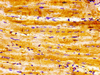
IHC image of SEMA3A Antibody diluted at 1:300 and staining in paraffin-embedded human heart tissue performed on a Leica BondTM system. After dewaxing and hydration, antigen retrieval was mediated by high pressure in a citrate buffer (pH 6.0). Section was blocked with 10% normal goat serum 30min at RT. Then primary antibody (1% BSA) was incubated at 4°C overnight. The primary is detected by a biotinylated secondary antibody and visualized using an HRP conjugated SP system.IHC
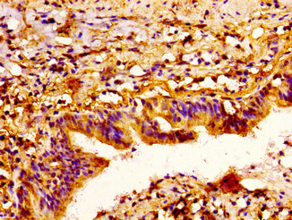
IHC image of SEMA3A Antibody diluted at 1:300 and staining in paraffin-embedded human lung cancer performed on a Leica BondTM system. After dewaxing and hydration, antigen retrieval was mediated by high pressure in a citrate buffer (pH 6.0). Section was blocked with 10% normal goat serum 30min at RT. Then primary antibody (1% BSA) was incubated at 4°C overnight. The primary is detected by a biotinylated secondary antibody and visualized using an HRP conjugated ABC system.| Product Name | SEMA3A Rabbit pAb |
|---|---|
| Antibody Type | Primary Antibodies |
| Immunogen | Recombinant Human Semaphorin-3A protein (357-445AA) |
| Clonality | polyclonal |
|---|---|
| Isotype | IgG |
| Host Species | Rabbit |
| Tested Applications | IHCWB |
| WB:1:500-1:5000 IHC:1:200-1:500 |
|
| Species Reactivity | HumanMouseRat |
| Concentration | 1mg/ml |
| Purification | Protein G |
| Gene Symbol | SEMA3A |
|---|---|
| Gene Synonyms | HH16 SemD COLL1 SEMA1 SEMAD SEMAL coll-1 Hsema-I SEMAIII Hsema-III |
| Gene Full Name | semaphorin 3A |
| Gene Summary | This gene is a member of the semaphorin family and encodes a protein with an Ig-like C2-type (immunoglobulin-like) domain, a PSI domain and a Sema domain. This secreted protein can function as either a chemorepulsive agent, inhibiting axonal outgrowth, or as a chemoattractive agent, stimulating the growth of apical dendrites. In both cases, the protein is vital for normal neuronal pattern development. Increased expression of this protein is associated with schizophrenia and is seen in a variety of human tumor cell lines. Also, aberrant release of this protein is associated with the progression of Alzheimer's disease. [provided by RefSeq, Jul 2008] |
| Molecular Weight(MW) | 89kDa |
| Cellular Localization | Secreted. |
WB
Western Blot Positive WB detected in: Hela whole cell lysate, 293 whole cell lysate, Rat kidney tissue, Mouse heart tissue, Mouse skeletal muscle tissue All lanes: SEMA3A antibody at 3.6µg/ml Secondary Goat polyclonal to rabbit IgG at 1/50000 dilution.
IHC
IHC image of SEMA3A Antibody diluted at 1:300 and staining in paraffin-embedded human heart tissue performed on a Leica BondTM system. After dewaxing and hydration, antigen retrieval was mediated by high pressure in a citrate buffer (pH 6.0). Section was blocked with 10% normal goat serum 30min at RT. Then primary antibody (1% BSA) was incubated at 4°C overnight. The primary is detected by a biotinylated secondary antibody and visualized using an HRP conjugated SP system.
IHC
IHC image of SEMA3A Antibody diluted at 1:300 and staining in paraffin-embedded human lung cancer performed on a Leica BondTM system. After dewaxing and hydration, antigen retrieval was mediated by high pressure in a citrate buffer (pH 6.0). Section was blocked with 10% normal goat serum 30min at RT. Then primary antibody (1% BSA) was incubated at 4°C overnight. The primary is detected by a biotinylated secondary antibody and visualized using an HRP conjugated ABC system.| Application Notes | WB:1:500-1:5000 IHC:1:200-1:500 |
|---|
| Form | Liquid |
|---|---|
| Storage Instructions | Upon receipt, store at -20°C or -80°C. Avoid repeated freeze. |
| Storage Buffer | Preservative: 0.03% Proclin 300,Constituents: 50% Glycerol, 0.01M PBS, pH 7.4 |


 Data sheet for OM644239