WB
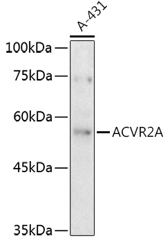
Western blot analysis of lysates from A-431 cells, using ACVR2A Rabbit pAb at 1:500 dilution. Secondary antibody: HRP-conjugated Goat anti-Rabbit IgG (H+L)at 1:10000 dilution. Lysates/proteins: 25μg per lane. Blocking buffer: 3% nonfat dry milk in TBST. Detection: ECL Enhanced Kit. Exposure time: 180s.IHC
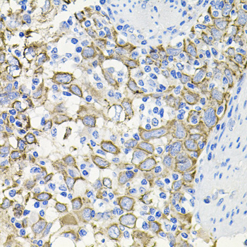
Immunohistochemistry analysis of paraffin embedded Human esophageal cancer using ACVR2A Rabbit pAb at dilution of 1:100 (40x lens). Microwave antigen retrieval performed with 0.01M PBS Buffer (pH 7.2) prior to IHC staining.IHC
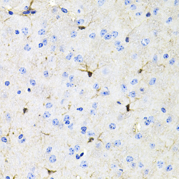
Immunohistochemistry analysis of paraffin embedded Mouse brain using ACVR2A Rabbit pAb at dilution of 1:100 (40x lens). Microwave antigen retrieval performed with 0.01M PBS Buffer (pH 7.2) prior to IHC staining.ICC/IF
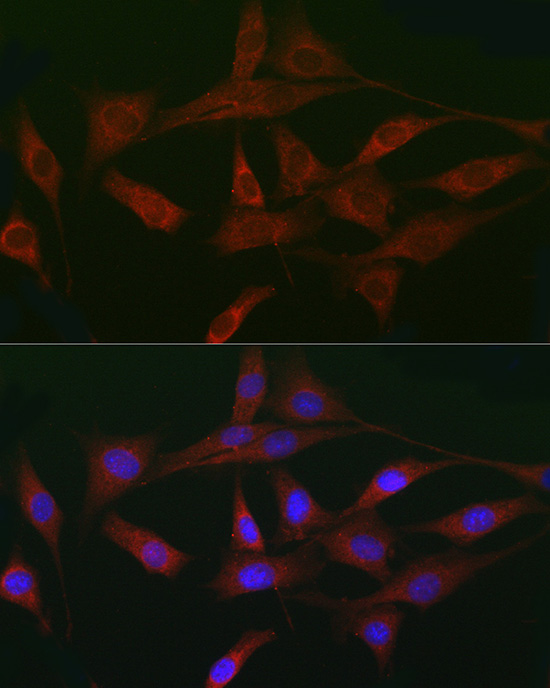
Immunofluorescence analysis of NIH/3T3 cells using ACVR2A Rabbit pAb at dilution of 1:50 (40x lens). Secondary antibody: Cy3-conjugated Goat anti-Rabbit IgG (H+L) at 1:500 dilution. Blue: DAPI for nuclear staining.| Product Name | ACVR2A Rabbit pAb |
|---|---|
| Antibody Type | Primary Antibodies |
| Immunogen | Recombinant fusion protein containing a sequence corresponding to amino acids 20-140 of human ACVR2A (NP_001607.1). |
| Clonality | Polyclonal |
|---|---|
| Isotype | IgG |
| Host Species | Rabbit |
| Tested Applications | ICC/IFIHCWB |
| WB:1:100-1:500 IHC:1:100-1:200 ICC/IF:1:50-1:200 |
|
| Species Reactivity | HumanMouseRat |
| Concentration | 1mg/ml |
| Purification | Affinity purified |
| Gene Symbol | ACVR2A |
|---|---|
| Gene Synonyms | ACVR2 ACTRII |
| Gene Full Name | activin A receptor type 2A |
| Gene Summary | This gene encodes a receptor that mediates the functions of activins, which are members of the transforming growth factor-beta (TGF-beta) superfamily involved in diverse biological processes. The encoded protein is a transmembrane serine-threonine kinase receptor which mediates signaling by forming heterodimeric complexes with various combinations of type I and type II receptors and ligands in a cell-specific manner. The encoded type II receptor is primarily involved in ligand-binding and includes an extracellular ligand-binding domain, a transmembrane domain and a cytoplasmic serine-threonine kinase domain. This gene may be associated with susceptibility to preeclampsia, a pregnancy-related disease which can result in maternal and fetal morbidity and mortality. Alternative splicing results in multiple transcript variants of this gene. [provided by RefSeq, Jun 2013] |
| Alternative Names | ACVR2 ACTRII ACVR2A |
| Molecular Weight(MW) | 58kDa |
| Cellular Localization | Membrane, Single-pass type I membrane protein. |
WB
Western blot analysis of lysates from A-431 cells, using ACVR2A Rabbit pAb at 1:500 dilution. Secondary antibody: HRP-conjugated Goat anti-Rabbit IgG (H+L)at 1:10000 dilution. Lysates/proteins: 25μg per lane. Blocking buffer: 3% nonfat dry milk in TBST. Detection: ECL Enhanced Kit. Exposure time: 180s.
IHC
Immunohistochemistry analysis of paraffin embedded Human esophageal cancer using ACVR2A Rabbit pAb at dilution of 1:100 (40x lens). Microwave antigen retrieval performed with 0.01M PBS Buffer (pH 7.2) prior to IHC staining.
IHC
Immunohistochemistry analysis of paraffin embedded Mouse brain using ACVR2A Rabbit pAb at dilution of 1:100 (40x lens). Microwave antigen retrieval performed with 0.01M PBS Buffer (pH 7.2) prior to IHC staining.
ICC/IF
Immunofluorescence analysis of NIH/3T3 cells using ACVR2A Rabbit pAb at dilution of 1:50 (40x lens). Secondary antibody: Cy3-conjugated Goat anti-Rabbit IgG (H+L) at 1:500 dilution. Blue: DAPI for nuclear staining.| Application Notes | WB:1:100-1:500 IHC:1:100-1:200 ICC/IF:1:50-1:200 |
|---|
| Form | Liquid |
|---|---|
| Storage Instructions | Store at -20℃. Avoid freeze / thaw cycles. |
| Storage Buffer | Buffer: PBS with 0.02% Sodium azide, 50% glycerol, pH7.3. |


