WB
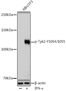
Western blot analysis of lysates from NIH/3T3 cells, using Phospho-Tyk2-Y1054/1055 Rabbit pAb at 1:1000 dilution. NIH/3T3 cells were treated by IFN-α (100 ng/ml) at 37℃ for 30 minutes after serum starvation overnight. Secondary antibody: HRP-conjugated Goat anti-Rabbit IgG (H+L) at 1:10000 dilution. Lysates/proteins: 25μg per lane. Blocking buffer: 3% BSA. Detection: ECL Enhanced Kit. Exposure time: 180s.| Product Name | Phospho-Tyk2-Y1054/1055 Rabbit pAb |
|---|---|
| Antibody Type | Primary Antibodies |
| Immunogen | A synthetic phosphorylated peptide around Y1054 & Y1055 of human Tyk2 (NP_003322.3). |
| Modification | p-Y1054/1055 |
| Clonality | Polyclonal |
|---|---|
| Isotype | IgG |
| Host Species | Rabbit |
| Tested Applications | WB |
| WB:1:500-1:1000 |
|
| Species Reactivity | HumanMouse |
| Concentration | 1mg/ml |
| Purification | Affinity purified |
| Gene Symbol | TYK2 |
|---|---|
| Gene Synonyms | JTK1 IMD35 |
| Gene Full Name | tyrosine kinase 2 |
| Gene Summary | This gene encodes a member of the tyrosine kinase and, more specifically, the Janus kinases (JAKs) protein families. This protein associates with the cytoplasmic domain of type I and type II cytokine receptors and promulgate cytokine signals by phosphorylating receptor subunits. It is also a component of both the type I and type III interferon signaling pathways. As such, it may play a role in anti-viral immunity. A mutation in this gene has been associated with Immunodeficiency 35. [provided by RefSeq, Sep 2020] |
| Alternative Names | JTK1 IMD35 Phospho-Tyk2-Y1054/1055 |
| Molecular Weight(MW) | 134kDa |
| Cellular Localization | cytoplasm, cytoskeleton, cytosol, extracellular exosome, nucleus. |
WB
Western blot analysis of lysates from NIH/3T3 cells, using Phospho-Tyk2-Y1054/1055 Rabbit pAb at 1:1000 dilution. NIH/3T3 cells were treated by IFN-α (100 ng/ml) at 37℃ for 30 minutes after serum starvation overnight. Secondary antibody: HRP-conjugated Goat anti-Rabbit IgG (H+L) at 1:10000 dilution. Lysates/proteins: 25μg per lane. Blocking buffer: 3% BSA. Detection: ECL Enhanced Kit. Exposure time: 180s.| Application Notes | WB:1:500-1:1000 |
|---|
| Form | Liquid |
|---|---|
| Storage Instructions | Store at -20℃. Avoid freeze / thaw cycles. |
| Storage Buffer | PBS with 0.02% sodium azide, 50% glycerol, pH7.3. |


