WB
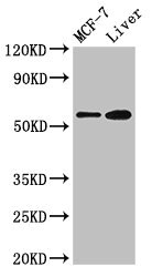
Positive WB detected in: MCF-7 whole cell lysate, Mouse liver tissue, All lanes: CBS antibody at 2.7µg/ml, Secondary Goat polyclonal to rabbit IgG at 1/50000 dilution.IHC
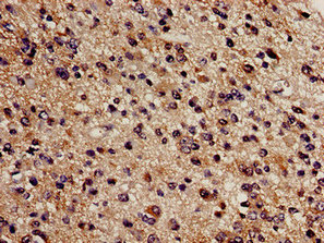
Immunohistochemistry analysis of human glioma using CBS Antibody at dilution of 1:100.IHC
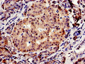
Immunocytochemistry analysis of human pancreatic tissue using CBS Antibody at dilution of 1:100.ICC/IF
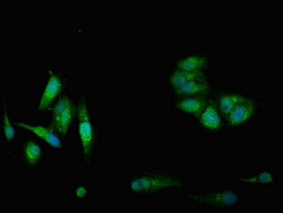
Immunofluorescent analysis of Hela cells using CBS Antibody at dilution of 1:100 and Alexa Fluor 488-congugated AffiniPure Goat Anti-Rabbit IgG(H+L).IP
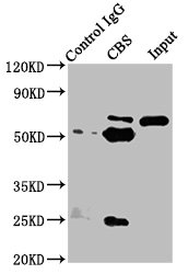
Immunoprecipitating CBS in Hela whole cell lysate, Lane 1: Rabbit control IgG instead of CBS Antibody in Hela whole cell lysate. For western blotting, a HRP-conjugated Protein G antibody was used as the secondary antibody (1/5000), Lane 2: CBS Antibody(8µg) + Hela whole cell lysate (500µg), Lane 3: Hela whole cell lysate (20µg).WB
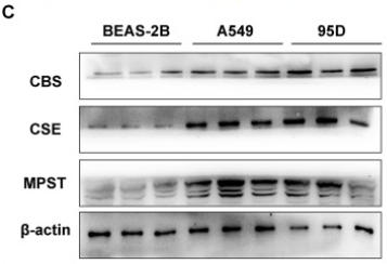
(C) Western blot analysis (left) and quantitative analysis (right) of CBS, CSE and MPST protein expression in lung cancer cell lines (A549 and 95D) and normal lung cell line (BEAS-2B).WB
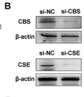
(B) Western blot analysis of CBS and CSE expressed in A549 cells after transfection.WB
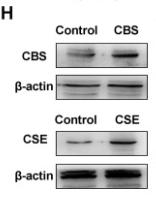
(H) Western blot analysis of CBS and CSE expressed in BEAS-2B cells after infection.WB
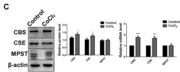
(C) Western blot analysis and qRT-PCR analysis of CBS, CSE and MPST expression after 200 μM CoCl2 treatment for 24 h.WB
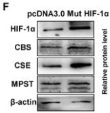
(F) Western blot and qRT-PCR analysis of H2S-producing enzymes and HIF-1α expression after transfection with pcDNA3.0/mut HIF-1α plasmid or empty vector control.WB
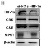
(H) The expression levels of CBS, CSE, MPST and HIF-1α in protein and mRNA were detected using western blot and qRT-PCR analysis after transfection with si-HIF-1α or si-negative control (si-NC), respectively.WB
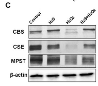
(C) Western blot analysis of CBS, CSE, and MPST protein expression levels.| Product Name | Rabbit anti-CBS Polyclonal antibody |
|---|---|
| Antibody Type | Primary Antibodies |
| Immunogen | Recombinant Human Cystathionine beta-synthase protein (1-413 AA). |
| Clonality | Polyclonal |
|---|---|
| Isotype | IgG |
| Host Species | Rabbit |
| Tested Applications | ICC/IFIHCIPWB |
| WB:1:500-1:5000 IHC:1:20-1:200 ICC/IF:1:50-1:200 IP:1:200-1:2000 |
|
| Species Reactivity | HumanMouse |
| Concentration | 1mg/ml |
| Purification | Protein G |
| Gene Symbol | CBS |
|---|---|
| Gene Synonyms | CBSL HIP4 |
| Gene Full Name | cystathionine beta-synthase |
| Gene Summary | The protein encoded by this gene acts as a homotetramer to catalyze the conversion of homocysteine to cystathionine, the first step in the transsulfuration pathway. The encoded protein is allosterically activated by adenosyl-methionine and uses pyridoxal phosphate as a cofactor. Defects in this gene can cause cystathionine beta-synthase deficiency (CBSD), which can lead to homocystinuria. This gene is a major contributor to cellular hydrogen sulfide production. Multiple alternatively spliced transcript variants have been found for this gene. [provided by RefSeq, Feb 2016] |
| Alternative Names | AI047524 antibody AI303044 antibody Beta thionase antibody Beta-thionase antibody Cbs antibody Cbs cystathionine beta-synthase antibody CBS_HUMAN antibody Cystathionine beta synthase antibody Cystathionine beta-synthase antibody EC 4.2.1.22 antibody HIP 4 antibody HIP4 antibody Methylcysteine synthase antibody MGC18856 antibody MGC18895 antibody MGC37300 antibody OTTHUMP00000109416 antibody OTTHUMP00000109418 antibody Serine sulfhydrase antibody |
| Molecular Weight(MW) | 61kDa |
| Cellular Localization | Cytoplasm,Nucleus. |
WB
Positive WB detected in: MCF-7 whole cell lysate, Mouse liver tissue, All lanes: CBS antibody at 2.7µg/ml, Secondary Goat polyclonal to rabbit IgG at 1/50000 dilution.
IHC
Immunohistochemistry analysis of human glioma using CBS Antibody at dilution of 1:100.
IHC
Immunocytochemistry analysis of human pancreatic tissue using CBS Antibody at dilution of 1:100.
ICC/IF
Immunofluorescent analysis of Hela cells using CBS Antibody at dilution of 1:100 and Alexa Fluor 488-congugated AffiniPure Goat Anti-Rabbit IgG(H+L).
IP
Immunoprecipitating CBS in Hela whole cell lysate, Lane 1: Rabbit control IgG instead of CBS Antibody in Hela whole cell lysate. For western blotting, a HRP-conjugated Protein G antibody was used as the secondary antibody (1/5000), Lane 2: CBS Antibody(8µg) + Hela whole cell lysate (500µg), Lane 3: Hela whole cell lysate (20µg).
WB
(C) Western blot analysis (left) and quantitative analysis (right) of CBS, CSE and MPST protein expression in lung cancer cell lines (A549 and 95D) and normal lung cell line (BEAS-2B).
WB
(B) Western blot analysis of CBS and CSE expressed in A549 cells after transfection.
WB
(H) Western blot analysis of CBS and CSE expressed in BEAS-2B cells after infection.
WB
(C) Western blot analysis and qRT-PCR analysis of CBS, CSE and MPST expression after 200 μM CoCl2 treatment for 24 h.
WB
(F) Western blot and qRT-PCR analysis of H2S-producing enzymes and HIF-1α expression after transfection with pcDNA3.0/mut HIF-1α plasmid or empty vector control.
WB
(H) The expression levels of CBS, CSE, MPST and HIF-1α in protein and mRNA were detected using western blot and qRT-PCR analysis after transfection with si-HIF-1α or si-negative control (si-NC), respectively.
WB
(C) Western blot analysis of CBS, CSE, and MPST protein expression levels.| Application Notes | WB:1:500-1:5000 IHC:1:20-1:200 ICC/IF:1:50-1:200 IP:1:200-1:2000 |
|---|
| Form | Liquid |
|---|---|
| Storage Instructions | Upon receipt, store at -20°C or -80°C. Avoid repeated freeze. |
| Storage Buffer | Preservative: 0.03% Proclin 300,Constituents: 50% Glycerol, 0.01M PBS, pH 7.4 |


