WB
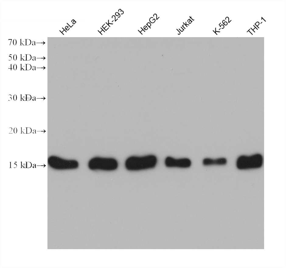
Various lysates were subjected to SDS PAGE followed by western blot with TOM20 antibody at dilution of 1:5000 incubated at room temperature for 1.5 hours.IHC
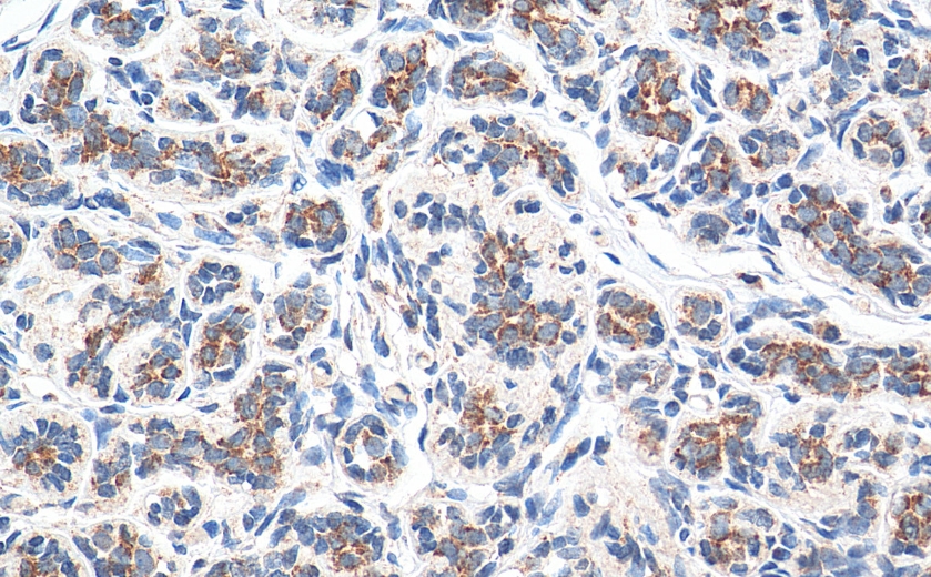
Immunohistochemical analysis of paraffin-embedded human breast cancer tissue slide using TOM20 antibody at dilution of 1:1000 (under 40x lens). Heat mediated antigen retrieval with Tris-EDTA buffer (pH 9.0).IHC
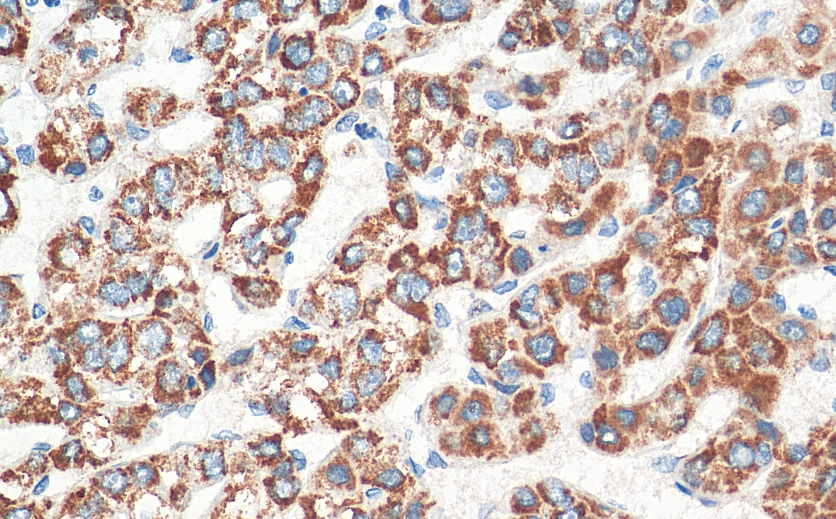
Immunohistochemical analysis of paraffin-embedded human liver cancer tissue slide using TOM20 antibody at dilution of 1:1000 (under 40x lens). Heat mediated antigen retrieval with Tris-EDTA buffer (pH 9.0).ICC/IF
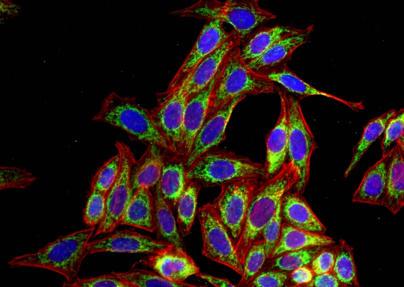
Immunofluorescent analysis of (4% PFA) fixed HepG2 cells using TOM20 antibody at dilution of 1:800 and Omnimabs® 488 Goat Anti-Mouse IgG(H&L), (CL594-Phalloidin, red).FC
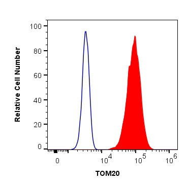
1X10^6 HeLa cells were intracellularly stained with 0.4 ug Anti-Human TOM20 and Omnimabs® 488 Goat Anti-Mouse IgG(H&L) at dilution 1:1000 (red), or 0.4 ug Control Antibody. Cells were fixed with 4% PFA and permeabilized with Flow Cytometry Perm Buffer.| Product Name | TOM20 Monoclonal antibody |
|---|---|
| Antibody Type | Primary Antibodies |
| Immunogen | Recombinant human TOM20 protein. |
| Clonality | Monoclonal |
|---|---|
| Isotype | IgG2b |
| Host Species | Mouse |
| Tested Applications | FCICC/IFIHCWB |
| WB:1:1000-1:5000 IHC:1:500-1:2000 ICC/IF:1:400-1:1600 FC:0.40 ug per 10^6 cells in a 100 µl suspension |
|
| Species Reactivity | Human |
| Concentration | 0.5mg/ml |
| Purification | Affinity purified |
| Gene Symbol | TOMM20 |
|---|---|
| Gene Synonyms | MAS20 MOM19 TOM20 |
| Gene Full Name | translocase of outer mitochondrial membrane 20 |
| Gene Summary | Enables mitochondrion targeting sequence binding activity; protein-transporting ATPase activity; and unfolded protein binding activity. Involved in protein targeting to mitochondrion. Located in mitochondria-associated endoplasmic reticulum membrane contact site and mitochondrial outer membrane. Is active in mitochondrion. [provided by Alliance of Genome Resources, Jun 2025] |
| Alternative Names | TOMM20 1D6F5 KIAA0016 MAS20 MOM19 |
| Molecular Weight(MW) | 16kDa |
| Cellular Localization | Mitochondrion outer membrane. |
WB
Various lysates were subjected to SDS PAGE followed by western blot with TOM20 antibody at dilution of 1:5000 incubated at room temperature for 1.5 hours.
IHC
Immunohistochemical analysis of paraffin-embedded human breast cancer tissue slide using TOM20 antibody at dilution of 1:1000 (under 40x lens). Heat mediated antigen retrieval with Tris-EDTA buffer (pH 9.0).
IHC
Immunohistochemical analysis of paraffin-embedded human liver cancer tissue slide using TOM20 antibody at dilution of 1:1000 (under 40x lens). Heat mediated antigen retrieval with Tris-EDTA buffer (pH 9.0).
ICC/IF
Immunofluorescent analysis of (4% PFA) fixed HepG2 cells using TOM20 antibody at dilution of 1:800 and Omnimabs® 488 Goat Anti-Mouse IgG(H&L), (CL594-Phalloidin, red).
FC
1X10^6 HeLa cells were intracellularly stained with 0.4 ug Anti-Human TOM20 and Omnimabs® 488 Goat Anti-Mouse IgG(H&L) at dilution 1:1000 (red), or 0.4 ug Control Antibody. Cells were fixed with 4% PFA and permeabilized with Flow Cytometry Perm Buffer.| Application Notes | WB:1:1000-1:5000 IHC:1:500-1:2000 ICC/IF:1:400-1:1600 FC:0.40 ug per 10^6 cells in a 100 µl suspension |
|---|
| Form | Liquid |
|---|---|
| Storage Instructions | Store at -20°C. Stable for one year after shipment. |
| Storage Buffer | PBS with 0.02% sodium azide and 50% glycerol pH 7.3. |


