WB
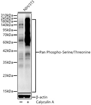
Western blot analysis of lysates from NIH/3T3 cells using Pan Phospho-Serine/Threonine Rabbit pAb at 1:400 dilution. NIH/3T3 cells were treated with Calyculin A (100 nM) at 37℃ for 30 minutes after serum starvation overnight. Secondary antibody: HRP-conjugated Goat anti-Rabbit IgG (H+L) at 1:10000 dilution. Lysates/proteins: 25 μg per lane. Blocking buffer: 3% nonfat dry milk in TBST. Detection: ECL Enhanced Kit. Exposure time: 45s.WB
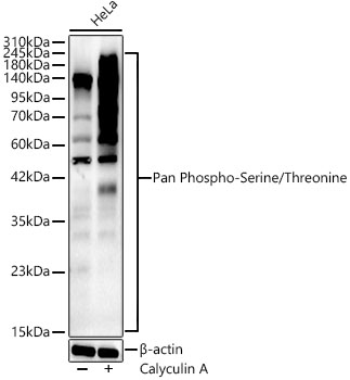
Western blot analysis of lysates from HeLa cells using Pan Phospho-Serine/Threonine Rabbit pAb at 1:400 dilution. HeLa cells were treated with Calyculin A (100 nM) at 37℃ for 30 minutes after serum starvation overnight. Secondary antibody: HRP-conjugated Goat anti-Rabbit IgG (H+L) at 1:10000 dilution. Lysates/proteins: 25 μg per lane. Blocking buffer: 3% nonfat dry milk in TBST. Detection: ECL Enhanced Kit. Exposure time: 45s.WB
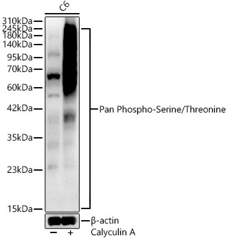
Western blot analysis of lysates from C6 cells using Pan Phospho-Serine/Threonine Rabbit pAb at 1:400 dilution. C6 cells were treated with Calyculin A (100 nM) at 37℃ for 30 minutes after serum starvation overnight. Secondary antibody: HRP-conjugated Goat anti-Rabbit IgG (H+L) at 1:10000 dilution. Lysates/proteins: 25 μg per lane. Blocking buffer: 3% nonfat dry milk in TBST. Detection: ECL Enhanced Kit. Exposure time: 45s.| Product Name | Pan Phospho-Serine/Threonine Rabbit pAb |
|---|---|
| Antibody Type | Primary Antibodies |
| Immunogen | Synthetic peptide. |
| Clonality | Polyclonal |
|---|---|
| Isotype | IgG |
| Host Species | Rabbit |
| Tested Applications | WB |
| WB:1:500-1:1000 |
|
| Species Reactivity | All |
| Concentration | 1mg/ml |
| Purification | Affinity purified |
| Molecular Weight(MW) | >10kDa |
|---|---|
| Function | As a critical post-translational modification, phosphorylation plays important roles in regulating various biological processes,Serine/threonine phosphorylation is an important mechanism that is involved in the regulation of protein function. Protein phosphorylation is the most well-studied post translational modification (PTM), in which a phosphoryl group from adenosine triphosphate (ATP) is covalently attached to a serine (~86%), threonine (~12%), or tyrosine (~2%) by a kinase and removed by a phosphatase. Phosphorylation at other amino acids have also been reported. Phosphorylation can modify protein structure, function, and interactions. As such, phosphorylation plays a critical role in virtually all cellular processes in homeostasis and disease, including signal transduction, cell cycle, differentiation, proliferation, metabolism, motility, and death. Importantly, phosphorylation at different residues can cause different outcomes. For example, RAF1 is a kinase central to the MAPK pathway that is activated when it is phosphorylated at serine (S) or threonine (T) residues S259, S338, S340/341, T491, or S494. However, phosphorylation at S289/296/301 results in the inhibition of RAF1 kinase activity. |
WB
Western blot analysis of lysates from NIH/3T3 cells using Pan Phospho-Serine/Threonine Rabbit pAb at 1:400 dilution. NIH/3T3 cells were treated with Calyculin A (100 nM) at 37℃ for 30 minutes after serum starvation overnight. Secondary antibody: HRP-conjugated Goat anti-Rabbit IgG (H+L) at 1:10000 dilution. Lysates/proteins: 25 μg per lane. Blocking buffer: 3% nonfat dry milk in TBST. Detection: ECL Enhanced Kit. Exposure time: 45s.
WB
Western blot analysis of lysates from HeLa cells using Pan Phospho-Serine/Threonine Rabbit pAb at 1:400 dilution. HeLa cells were treated with Calyculin A (100 nM) at 37℃ for 30 minutes after serum starvation overnight. Secondary antibody: HRP-conjugated Goat anti-Rabbit IgG (H+L) at 1:10000 dilution. Lysates/proteins: 25 μg per lane. Blocking buffer: 3% nonfat dry milk in TBST. Detection: ECL Enhanced Kit. Exposure time: 45s.
WB
Western blot analysis of lysates from C6 cells using Pan Phospho-Serine/Threonine Rabbit pAb at 1:400 dilution. C6 cells were treated with Calyculin A (100 nM) at 37℃ for 30 minutes after serum starvation overnight. Secondary antibody: HRP-conjugated Goat anti-Rabbit IgG (H+L) at 1:10000 dilution. Lysates/proteins: 25 μg per lane. Blocking buffer: 3% nonfat dry milk in TBST. Detection: ECL Enhanced Kit. Exposure time: 45s.| Application Notes | WB:1:500-1:1000 |
|---|
| Form | Liquid |
|---|---|
| Storage Instructions | Store at -20℃. Avoid freeze / thaw cycles. |
| Storage Buffer | PBS with 0.05% proclin300, 50% glycerol, pH7.3. |


