WB
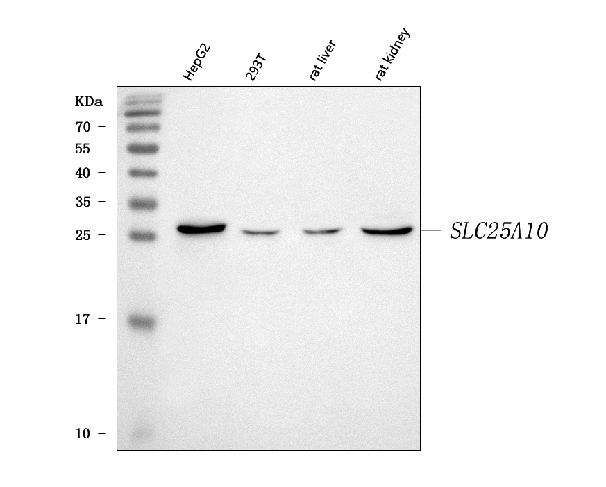
Western blot analysis of anti- Mitochondrial dicarboxylate carrier/SLC25A10 antibody. The sample well of each lane was loaded with 30ug of sample under reducing conditions. Lane 1: human HepG2 whole cell lysates, Lane 2: human 293T whole cell lysates, Lane 3: rat liver tissue lysates, Lane 4: rat kidney tissue lysates. Use rabbit anti- Mitochondrial dicarboxylate carrier/SLC25A10 1:1000, probed with a goat anti-rabbit IgG-HRP secondary antibody. The signal is developed using an Enhanced Chemiluminescent detection (ECL) kit.IHC
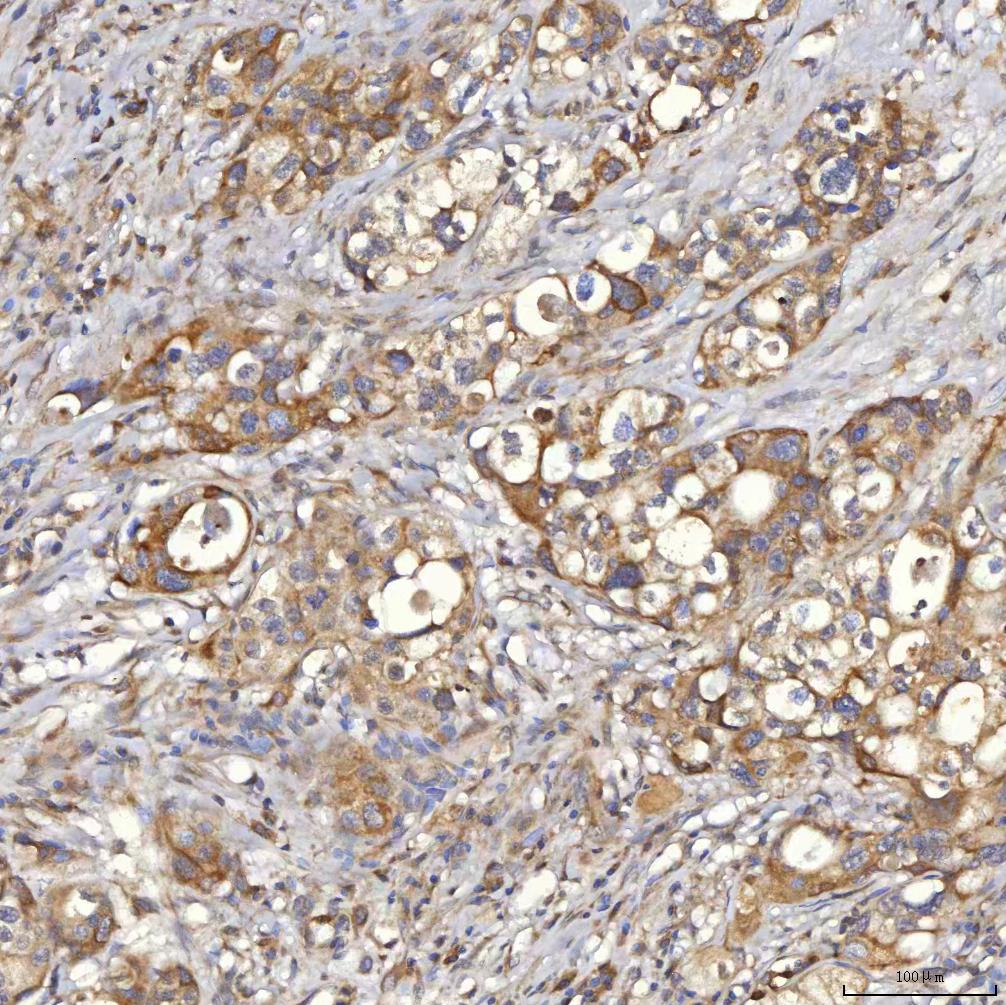
IHC analysis of SLC25A10 using anti-SLC25A10 antibody. SLC25A10 was detected in a paraffin-embedded section of human appendix adenocarcinoma tissue. The tissue section was incubated with rabbit anti-SLC25A10 Antibody at a dilution of 1:200 and developed using HRP Conjugated Rabbit IgG Super Vision Assay Kit with DAB as the chromogen.IHC
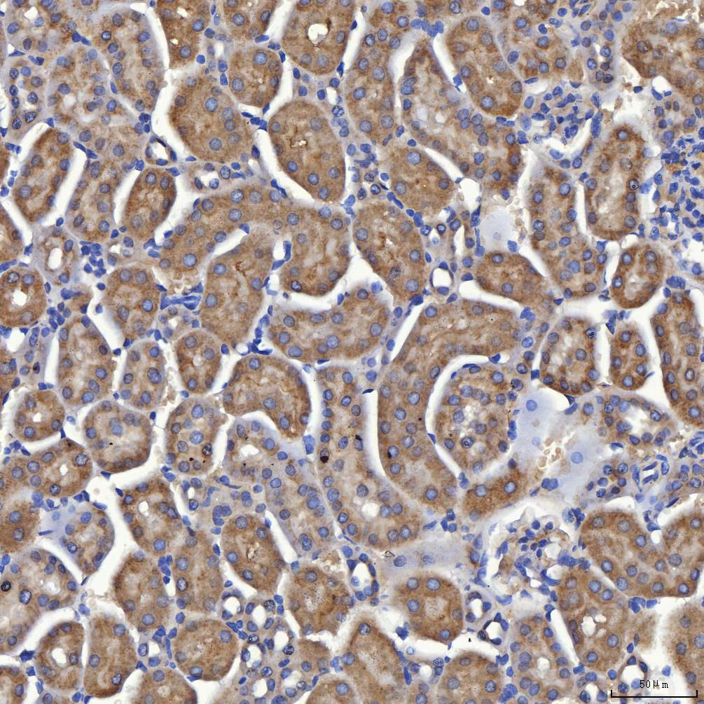
IHC analysis of SLC25A10 using anti-SLC25A10 antibody. SLC25A10 was detected in a paraffin-embedded section of mouse kidney tissue. The tissue section was incubated with rabbit anti-SLC25A10 Antibody at a dilution of 1:200 and developed using HRP Conjugated Rabbit IgG Super Vision Assay Kit with DAB as the chromogen.IHC
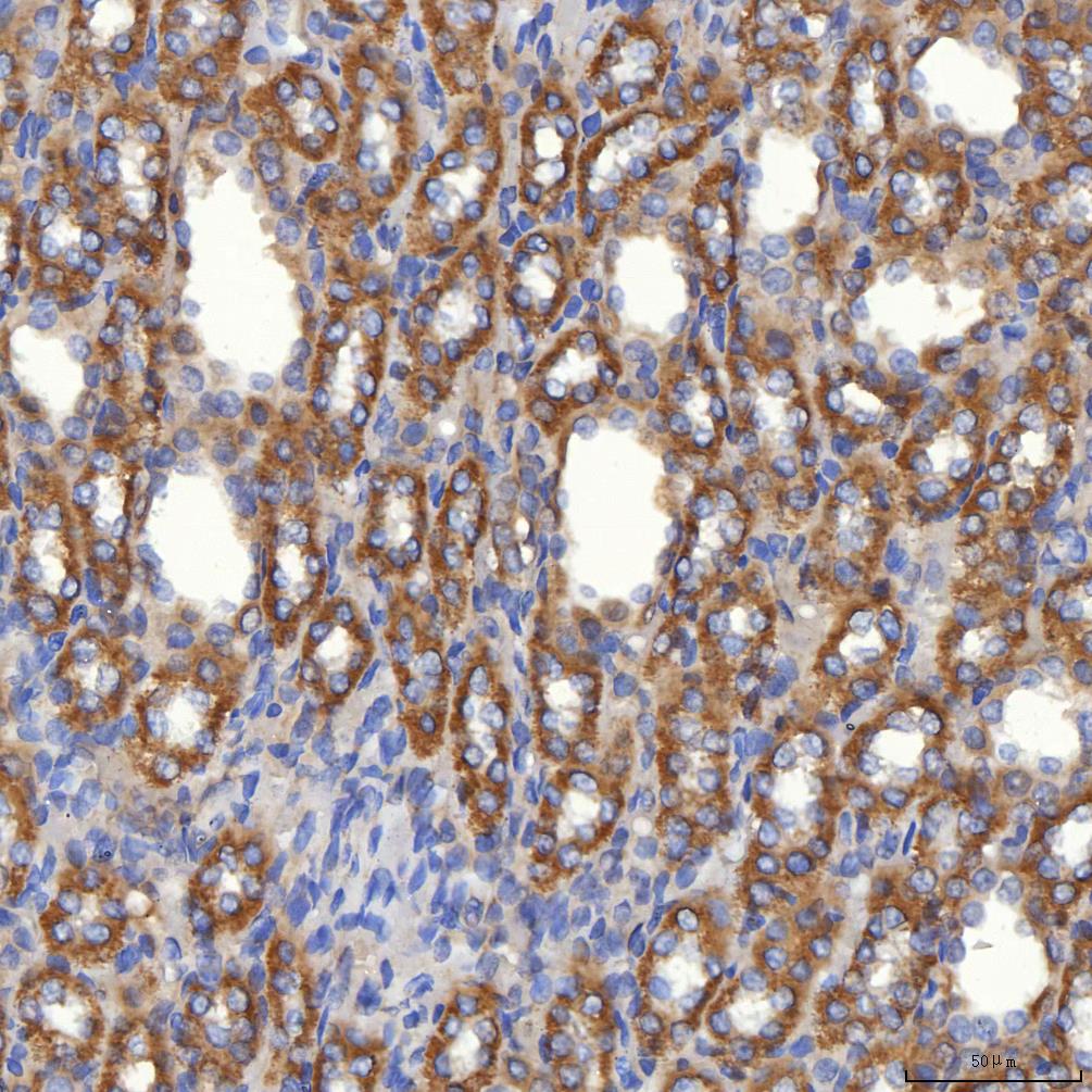
IHC analysis of SLC25A10 using anti-SLC25A10 antibody. SLC25A10 was detected in a paraffin-embedded section of rat kidney tissue. The tissue section was incubated with rabbit anti-SLC25A10 Antibody at a dilution of 1:200 and developed using HRP Conjugated Rabbit IgG Super Vision Assay Kit with DAB as the chromogen.IF-P
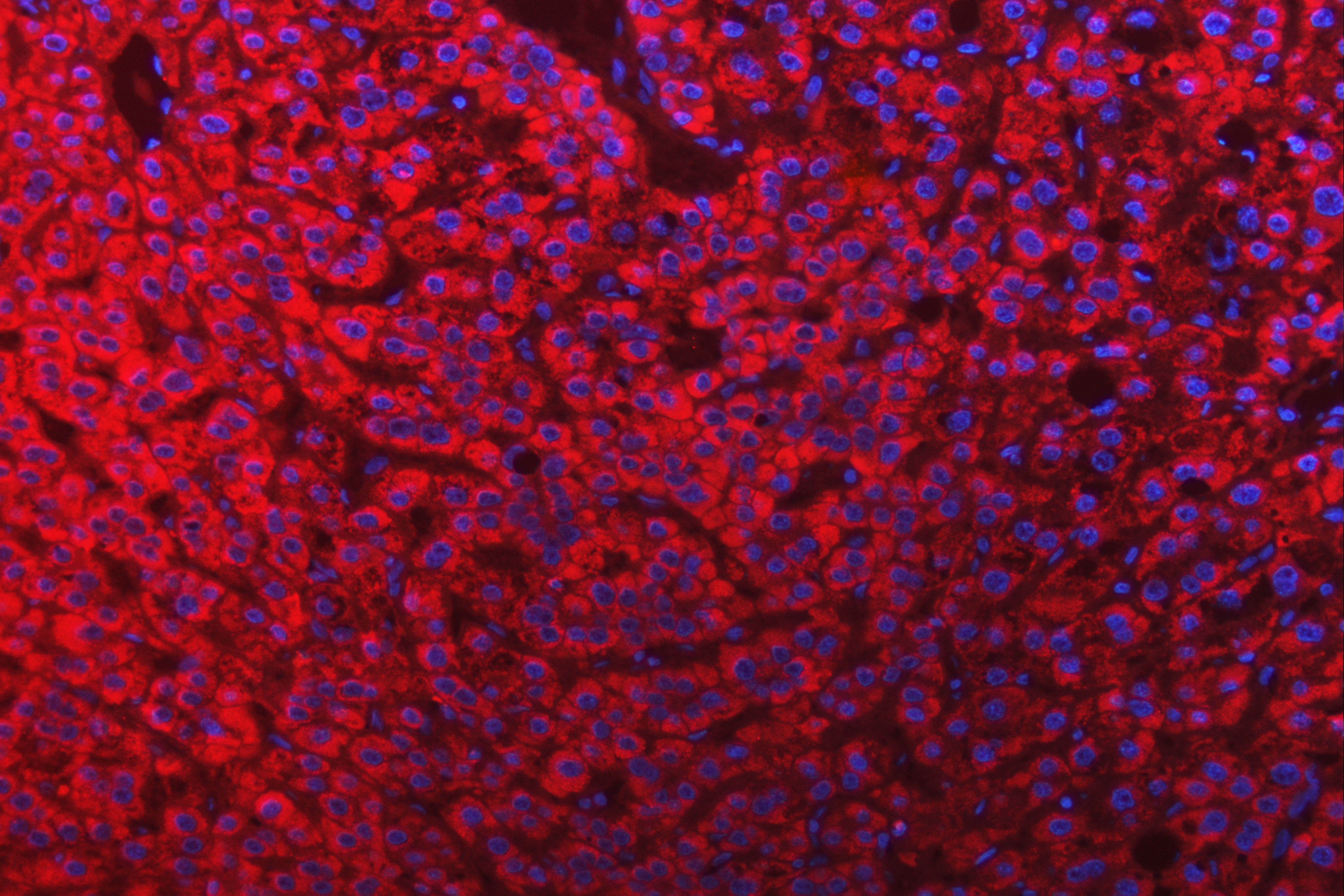
IF analysis using anti- Mitochondrial dicarboxylate carrier/SLC25A10 antibody. detected in paraffin-embedded section of human liver cancer tissue. The tissue section were stained using the cy3-conjugated Anti-rabbit IgG Secondary Antibody (red) and counterstained with DAPI (blue).| Product Name | Rabbit polyclonal antibody to SLC25A10 |
|---|---|
| Antibody Type | Primary Antibodies |
| Immunogen | E.coli-derived human Mitochondrial dicarboxylate carrier/SLC25A10 recombinant protein (Position: Q37-Q276). |
| Clonality | Polyclonal |
|---|---|
| Isotype | IgG |
| Host Species | Rabbit |
| Tested Applications | IF-PIHCWB |
| WB:1:500-1:2000 IHC:1:50-1:400 ICC/IFIF-P:1:50-1:400 |
|
| Species Reactivity | HumanMouseRat |
| Concentration | 0.5mg/ml |
| Purification | Affinity purified |
| Gene Symbol | SLC25A10 |
|---|---|
| Gene Synonyms | DIC MTDPS19 |
| Gene Full Name | solute carrier family 25 member 10 |
| Gene Summary | This gene encodes a member of a family of proteins that translocate small metabolites across the mitochondrial membrane. The encoded protein exchanges dicarboxylates, such as malate and succinate, for phosphate, sulfate, and other small molecules, thereby providing substrates for metabolic processes including the Krebs cycle and fatty acid synthesis. Alternatively spliced transcript variants encoding multiple isoforms have been observed for this gene. [provided by RefSeq, Aug 2012] |
| Alternative Names | DIC SLC25A10 |
| Molecular Weight(MW) | 31kDa |
| Cellular Localization | Mitochondrion inner membrane. |
WB
Western blot analysis of anti- Mitochondrial dicarboxylate carrier/SLC25A10 antibody. The sample well of each lane was loaded with 30ug of sample under reducing conditions. Lane 1: human HepG2 whole cell lysates, Lane 2: human 293T whole cell lysates, Lane 3: rat liver tissue lysates, Lane 4: rat kidney tissue lysates. Use rabbit anti- Mitochondrial dicarboxylate carrier/SLC25A10 1:1000, probed with a goat anti-rabbit IgG-HRP secondary antibody. The signal is developed using an Enhanced Chemiluminescent detection (ECL) kit.
IHC
IHC analysis of SLC25A10 using anti-SLC25A10 antibody. SLC25A10 was detected in a paraffin-embedded section of human appendix adenocarcinoma tissue. The tissue section was incubated with rabbit anti-SLC25A10 Antibody at a dilution of 1:200 and developed using HRP Conjugated Rabbit IgG Super Vision Assay Kit with DAB as the chromogen.
IHC
IHC analysis of SLC25A10 using anti-SLC25A10 antibody. SLC25A10 was detected in a paraffin-embedded section of mouse kidney tissue. The tissue section was incubated with rabbit anti-SLC25A10 Antibody at a dilution of 1:200 and developed using HRP Conjugated Rabbit IgG Super Vision Assay Kit with DAB as the chromogen.
IHC
IHC analysis of SLC25A10 using anti-SLC25A10 antibody. SLC25A10 was detected in a paraffin-embedded section of rat kidney tissue. The tissue section was incubated with rabbit anti-SLC25A10 Antibody at a dilution of 1:200 and developed using HRP Conjugated Rabbit IgG Super Vision Assay Kit with DAB as the chromogen.
IF-P
IF analysis using anti- Mitochondrial dicarboxylate carrier/SLC25A10 antibody. detected in paraffin-embedded section of human liver cancer tissue. The tissue section were stained using the cy3-conjugated Anti-rabbit IgG Secondary Antibody (red) and counterstained with DAPI (blue).| Application Notes | WB:1:500-1:2000 IHC:1:50-1:400 ICC/IFIF-P:1:50-1:400 |
|---|
| Form | Liquid |
|---|---|
| Storage Instructions | 12 months from date of receipt, -20℃ as supplied. 6 months 2 to 8℃ after reconstitution. Avoid repeated freezing and thawing. |
| Storage Buffer | 500ug/ml antibody with PBS, 0.02% NaN3, 1 mg/ml BSA and 50% glycerol. |


